As splenomegaly doctors refer to an enlarged spleen. It can be a symptom of various illnesses such as infections, blood disorders or liver damage. Patients occasionally complain of a feeling of pressure in the left upper abdomen. The therapy depends on the underlying disease. Eventually, the spleen must be removed. Read all about splenomegaly here!
Splenomegaly: description
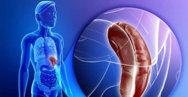
An enlarged spleen is a common symptom. It can occur in different diseases. These include infectious diseases, hereditary diseases, diseases of the blood or liver and many more. To better understand what the tasks and location of the spleen are in the body, let’s take a look at the anatomy and physiology of the spleen.
Splenomegaly: anatomy and physiology of the spleen
The spleen is also referred to as Splen or Lien. As the body’s largest lymphatic organ, it is an important part of the immune system. It lies in the left upper abdomen, usually behind the lower ribs. It is usually about four inches thick, seven inches wide and eleven inches long. It is enveloped by a capsule. Their tasks include trapping and breaking down old and deformed blood cells as well as microorganisms found in the blood. In addition, immune cells mature in her. It is possible to live without spleen. However, then increases the risk of serious infections.
Splenomegaly: symptoms
Splenomegaly is itself a disease symptom, but in turn causes other complaints. Therefore, in splenomegaly one has to distinguish symptoms caused by spleen enlargement itself and those caused by the underlying disease.
Splenomegaly: symptoms of the underlying disease
Many different diseases can lead to splenomegaly. Depending on this underlying disease, the patients have complaints. As a guide, the following relationships can be assumed:
- In infectious diseases: fever, fatigue, lymphadenopathy
- For malignant diseases: weight loss, night sweats, fever
- In blood disorders: fatigue, weakness, paleness
- In liver damage: jaundice, esophageal bleeding, visible abdominal veins
Spleen enlargement: Symptoms that causes it
A morbid spleen swelling is usually palpable under the left costal arch. It can cause pain, for example when it presses on nerves or displaces other organs. If the spleen swells too much for the capsule that surrounds it, it can rupture. The so-called splenic rupture is accompanied by severe pain in the left upper abdomen. This pain can radiate into the left shoulder.
Splenomegaly: causes and risk factors
The causes that can lead to splenomegaly are manifold. They can be divided into different groups.
blood diseases
There are benign and malignant diseases of the blood that can lead to splenomegaly. Benign ones include congenital red blood cell defects. These are:
- Sickle cell anemia
- thalassemia
- Hereditary spherocytosis
- Glucose-6-phosphate dehydrogenase deficiency
These diseases lead to a change in the erythrocyte structure. These remain in the vessels of the spleen and are degraded there. Since many erythrocytes accumulate, the spleen becomes larger to accomplish the numerous degradation.
Malignant diseases of the blood that cause an enlarged spleen are leukemias and lymphomas as well as myeloproliferative disorders such as osteomyelofibrosis or juvenile myelomonocytic leukemia.
infections
Typical for spleen enlargement is an infection with Epstein-Barr virus (EBV), which triggers mononucleosis (glandular fever). Cytomegalovirus (CMV) also causes splenomegaly in children. Other infectious diseases that may be associated with splenomegaly include:
- bacterial sepsis
- leishmaniasis
- malaria
- syphilis
- typhus
- tuberculosis
Pfortaderschädigungen
If there is a drainage obstruction in the portal vein, the blood builds up in the spleen (congestion spleen). Reasons can be:
- heart failure
- Cirrhosis or fibrosis
- Portal vein thrombosis
- Budd-Chiari syndrome
storage diseases
Diseases associated with a metabolic disorder can lead to spleen enlargement. These include:
- Glycogen
- Morbus Niemann-Pick
- Gaucher’s disease
- mucopolysaccharidosis
Immunological diseases
Various immunological diseases can be the cause of splenomegaly. These include:
- Chediak-Higashi syndrome
- Kawasaki disease
- histiocytoses
- Chronic granulomatosis
- Autoimmune lymphoproliferative syndrome (ALPS)
Other causes
Other causes of an enlarged spleen may be space demands in the organ itself. These include distant metastases of malignant tumors, hemangiomas or hamartomas of the spleen. Lymphomas and leukemias can also infiltrate the spleen tissue.
In rare cases, spleen swelling may also occur in collagenoses such as systemic lupus erythematosus, Still’s disease or juvenile rheumatoid arthritis. Even with sarcoidosis splenomegaly is possible.
Splenomegaly: examinations and diagnosis
If you feel ill and feel pain in your left upper abdomen, visit your family doctor. He can diagnose splenomegaly and order further investigations on the causes. First, he will ask you in detail about your medical history (anamnesis). He may ask you the following questions:
- Have you been suffering from an infection lately?
- Are you suffering from a chronic or malignant disease?
- Do you have fever?
- Have you lost weight unwillingly lately?
- Do you wake up sweat-drenched at night?
Splenomegaly: physical examination
A large spleen can be palpated during physical examination under the left rib cage. The patient lies on the right side. The examiner stabilizes the patient with his left hand while he scans the left upper abdomen with his right hand. Usually the spleen should not be palpable. If your doctor feels it, you have splenomegaly. He can then confirm this in an ultrasound examination in which he measures the spleen. In addition, he can find in ultrasound evidence of liver damage or disease of the portal vein.
In addition, her doctor looks for swollen lymph nodes and signs of liver damage during physical examination. This can be a yellowing of the skin, redness on the palms, visible veins of the abdominal wall or small, star-like sponges on the décolleté.
Splenomegaly: advanced diagnostics
If your doctor has detected splenomegaly, further investigation is needed to find causes for the enlarged spleen. First, blood should be taken from the patient. In the laboratory it is examined:
- Blood count and blood smear (number of red and white blood cells and platelets including listing of different types of white blood cells and number of young red blood cells)
- Erythrocyte sedimentation rate
- Indications of liver damage: transaminases (ALAT, ASAT), bilirubin
- Immune parameters (C-reactive protein, antinuclear antibodies, rheumatoid factors, Coombs test, electrophoresis)
- Sign for viral infections
According to the results of the blood test, certain diseases may be excluded as the cause of splenomegaly, while others may be considered probable causes. Subsequently, further diagnostic steps can be initiated, such as an X-ray of the thorax, a computed tomography of the abdomen or a bone marrow biopsy.
Splenomegaly: treatment
Splenomegaly is usually a symptom of another underlying disease. Accordingly, after diagnosis of the underlying disease this must be treated. With effective therapy, splenomegaly often disappears.
The treatment of splenomegaly also involves surgical removal of the spleen (splenectomy). This is necessary if the spleen capsule ruptures due to the increase in size or an overfunction of the spleen (hypersplenism) arises. Splenectomy should be used as a last resort, as it carries the risk of serious infectious diseases. This is referred to as overwhelming post-splenectomy (OPSI) syndrome. Above all, encapsulated bacteria such as pneumococci or meningococci can no longer be sufficiently warded off.
Splenomegaly: disease course and prognosis
The disease process and prognosis of splenomegaly depend strongly on the underlying disease that causes the swollen spleen. Without treatment of the cause, the spleen may swell so much that its capsule ruptures. Depending on the severity of the tear, surgery may be required. If haemostasis is successful, surgery can be performed to preserve the condition. If the bleeding can not be stopped, the spleen must be removed immediately.
Another complication that can lead to splenectomy is the so-called hypersplenism. It is an over-function of the spleen. It then removes more blood cells than necessary (excessive phagocytosis). Since the bone marrow can not produce the missing blood cells fast enough, pancytopenia occurs. By this medical experts understand that there are too few red and white blood cells as well as platelets in the blood. This leads to weakness, susceptibility to infection and diffuse bleeding. In this case should be at splenomegaly the spleen are removed.