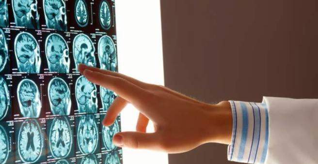
Cerebral haemorrhage (brain haemorrhage) occurs when a blood vessel bursts in the brain. Doctors also speak of a stroke caused by cerebral hemorrhage (haemorrhagic cerebral infarction). The escaping blood can disturb the function in the affected brain area. For example, intracerebral hemorrhage (ICB), subarachnoid haemorrhage, and epidural haemorrhage are distinguished depending on where the cerebral hemorrhage occurs. Read more about causes, diagnosis and treatment of cerebral hemorrhage.

Cerebral haemorrhage: description
In a stroke caused by cerebral hemorrhage (haemorrhagic stroke or infarction) forms a bruise (hematoma) in the brain tissue, which results from the bursting of a blood vessel. It comes to dysfunction in the affected area, and the brain tissue dies partially.
The umbrella term cerebral hemorrhage encompasses various clinical pictures:
Intracerebral haemorrhage (parenchymatous haemorrhage, intracerebral hematoma)
An intracerebral hemorrhage (ICB) is a hemorrhage into the brain tissue (brain parenchyma), caused by the bursting of a cerebral vasculature. A major risk factor for this is hypertension, especially in combination with arteriosclerosis. Because intracerebral haemorrhage usually affects a relatively large area of the brain, physicians also speak of brain mass haemorrhage. The term “intracerebral hematoma” is also used more often. After the exact location of the hematoma, this form of cerebral hemorrhage is subdivided further, for example into supra- or infratentorial hematomas. These are hematomas above or below the tentorium (small brain tent, a skin between the cerebellum and the cerebrum).
The intracerebral hemorrhage accounts for about ten percent of all strokes. The remaining 90 percent of the cases involve a stroke due to vascular occlusion (ischemic cerebral infarction).
Epidural haemorrhage (epidural haemorrhage, epidural hematoma)
In an epidural hemorrhage, blood accumulates between the skull bone and the outermost meninges (hard meninges or dura mater). This form of cerebral hemorrhage is usually traumatic and usually occurs in conjunction with a skull fracture.
Subdural hemorrhage (subdural haemorrhage, subdural hematoma)
In subdural haemorrhage, blood collects between the dura mater and the arachnoid membrane. Again, the cause is usually a traumatic crack of blood vessels. In some cases the subdural hematoma develops rapidly / acutely, in other cases slowly / chronically. Overall, subdural hemorrhage is three to five times more common than epidural hemorrhage.
subarachnoid hemorrhage
Subarachnoid haemorrhage develops directly on the brain surface and can lead to strokes. Read all about it in the article Subarachnoidal Hemorrhage!
Cerebral haemorrhage: symptoms
You can read all about possible signs of cerebral hemorrhage in the article Cerebral Hemorrhage – Symptoms.
Cerebral haemorrhage: causes and risk factors
high blood pressure
The most common cause of stroke due to cerebral hemorrhage (haemorrhagic cerebral infarction) is high blood pressure (hypertension). It causes the vessel walls to lose their elasticity over the years. Sudden spikes in blood pressure (for example, during exercise or a hypertensive crisis) can then easily rupture the vessels in the brain and cause cerebral hemorrhage.
Other causes
Ischemic strokes (cerebral infarction due to vascular occlusion) may bleed in their course.
Cerebral haemorrhage can also be caused by vascular malformations, such as sagging of the vessel wall (aneurysms, aneurysms). Another possible cause of cerebral hemorrhage is abnormally brittle vessels, such as amyloid angiopathy – a vascular disease whose cause is not well known. In the process, abnormally altered proteins (amyloids) are deposited in the vessel walls, which makes the vessels fragile. This can repeatedly lead to a brain hemorrhage. In the long term, these patients often suffer from mental deterioration (dementia).
In some cases, cerebral hemorrhage is a consequence of an increased bleeding tendency (bleeding diathesis). Such is, for example, when taking anticoagulant drugs, hemophilia, platelet deficiency (thrombocytopenia) and leukemia.
Cerebral haemorrhage can also occur in connection with substance abuse (cocaine, amphetamines, etc.), traumatic brain injury and tumors.
Cerebral haemorrhage: examinations and diagnosis
Rapid diagnosis and rapid onset of therapy are very important for cerebral hemorrhage.
Consult and examine
The diagnosis of stroke due to cerebral hemorrhage begins with a neurological examination. It examines the patient’s state of consciousness and the function of various nerves. The temporal development and accompanying circumstances of the complaints are important for the doctor. However, since people with a fresh cerebral hemorrhage are often unfocused and can not provide accurate information, a companion should be on site and describe to the doctor the Hergang.
By means of computer tomography (CT) of the head, a cerebral hemorrhage can be recognized immediately after its occurrence: The doctor sees the leaked blood as a “bright spot” and can determine the exact position and extent of the cerebral hemorrhage. In addition, on CT, a stroke due to cerebral hemorrhage (hemorrhagic stroke) can be distinguished from a stroke due to vascular occlusion (ischemic stroke) – both often cause very similar symptoms.
An alternative to CT is MRI (Magnetic Resonance Imaging, MRI). Here, too, a cerebral hemorrhage shows a patchy change in the brain.
Further investigations
Sometimes, for accurate diagnosis of stroke due to cerebral hemorrhage, further investigation is needed to visualize vascular malformations (for example, congenital saccule = aneurysms) or vascular leakage in the brain. Here angiography (X-ray examination of the vessels) is used, often in combination with magnetic resonance imaging (MR angiography) or computed tomography (CT angiography).
On the basis of blood tests, the doctors can detect, for example, coagulation disorders as a cause of cerebral hemorrhage. The causes of reduced blood clotting include hemophilia, the use of blood-thinning medications and advanced liver disease (liver cirrhosis).
Cerebral haemorrhage: treatment
In the hospital
Patients with a stroke due to cerebral hemorrhage must always be treated in the clinic. In the case of a major cerebral hemorrhage, brain swelling (edema) develops in the immediate vicinity, and the pressure in the skull can rise sharply. Then, for example, the following therapeutic measures can be initiated:
- Administration of dehydrating infusions
- artificial respiration
- Drug treatment with lower pressure increase
- Surgery to remove the bruise in case of strong pressure increase
- drug lowering of blood pressure
In cases of subarachnoid haemorrhage or hemorrhages in the brain’s interior, which are filled with cerebral water (ventricles), cerebral congestion (hydrocephalus) can develop. He can also be treated surgically, the ventricles receive a derivative (shunt).
rehabilitation
If the critical phase is over after a stroke due to cerebral hemorrhage after a few days, the early rehabilitation begins immediately. The aim is to enable patients to return to their current social environment and working life as quickly as possible.
Patients are cared for by a rehabilitation team consisting of physiotherapists, speech and occupational therapists as well as nurses and nurses. The relatives can also support the patient in many situations. The sooner the rehabilitation after a stroke starts due to cerebral hemorrhage (or vascular occlusion), the better the chances of success. Whether a long-term rehabilitation is required, depends on the extent of the nerve damage suffered as a result of cerebral hemorrhage.
Consequences of hemorrhage in the brain are often movement disorders. Through appropriate training routines such as Bobath, Vojta, Perfetti and Forced Use, therapists try to reverse as much as possible during physical rehabilitation.
An attempt is also made to improve speech disorders (aphasia), speech (dysarthria), vision, memory and attention. Some patients quickly recover completely after cerebral hemorrhaging, while others take months to years to regain control over their day-to-day activities.
In addition, the autonomy of stroke patients is promoted in rehab. Some sufferers have to learn to wash themselves, get dressed or cook.
Another goal in the treatment of cerebral hemorrhage is to lower blood pressure consistently and to normal levels over the long term.
Sometimes physical functions impaired by cerebral hemorrhage can not be improved despite intensive rehabilitation. Then it says: Develop strategies together with the patient, so that he can cope better with the restrictions. In dysphagia, for example, special body and head postures are practiced to prevent ingestion.
Cerebral haemorrhage: prevention
Congenital vascular malformations, which can lead to cerebral hemorrhage, can not be prevented.
The situation is different for hypertension, another risk factor for a stroke caused by cerebral hemorrhage. He should be treated accordingly. It is important that high blood pressure patients regularly measure their blood pressure and consistently take the prescribed medication – even if they are doing just fine. In addition to medication, regular exercise (at least 30 minutes several times a week) and a normal weight can reduce the blood pressure measurably.
Smokers have a two to three times higher risk of cerebral hemorrhage. Overweight, regular, high alcohol consumption and high cholesterol additionally increase the risk. Therefore, try to stop smoking, drink only moderately alcohol, incorporate more exercise into your daily routine, and respect a healthy diet (daily fresh fruits and vegetables, etc.). A conscious diet and exercise help obese people to lose weight – even a few kilos less bowing of a cerebral hemorrhage and many other health problems.
Cerebral haemorrhage: disease course and prognosis
The course of the first weeks after a stroke due to cerebral hemorrhage is very important for the prognosis. Doctors can estimate the further course by certain factors. These include the size of the cerebral hemorrhage, the spread of the blood into the brain chambers (ventricles), the age and the state of consciousness with which the patient was admitted to the hospital after the stroke. Overall, the long-term prognosis is after one cerebral hemorrhage rather unfavorable.