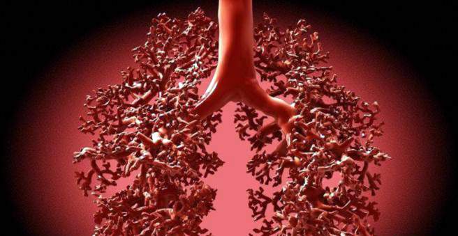
Bronchiectasis is a saccular enlargement of the bronchi in the lungs that can not regress. Bronchiectasis may have both congenital and acquired causes. Typical for bronchiectasis is a strong cough with a lot of mucous expectoration. If left untreated, bronchiectasis can lead to severe damage to the lungs. Learn about the causes, symptoms and treatment of bronchiectasis.

Bronchiectasis: description
Bronchiectasis (Greek: éktasis “expansion”) refers to pathological enlargements of the bronchi in the lungs. The condition when many bronchi are dilated in this way is called bronchiectasis. Various causes lead to damage to the bronchial walls, which ultimately results in a permanent expansion of the bronchi. Through targeted antibiotic therapy and vaccinations, bronchiectasis is less common in Germany today than it used to be. However, the high-resolution computed tomography (HR-CT) images reveal bronchiectasis more frequently and earlier, leading to an apparent increase in cases of illness.
How do bronchiectases develop?
The bronchi are the airways in the lungs. With every breath, inhaled air flows through them into the pulmonary alveoli, where the gas exchange takes place. With the respiratory air but also pathogens and dirt particles get into the bronchial system, which in the healthy by a sophisticated self-cleaning mechanism (the so-called mucociliary clearance) are continuously transported outside again. Namely, the airways are lined with certain cells that produce a mucous secretion and on whose surface the finest cilia (cilia) sit. The secretion helps to kill pathogens. The cilia constantly carry out a fanning movement toward the mouth (ciliary beat), through which the mucus and the attached pathogens and foreign bodies are transported to the throat. Once there, they are swallowed or coughed off.
This self-cleaning mechanism is important to keep the lungs free of foreign bodies and to prevent respiratory infections. The mechanism may be disturbed by various causes, which can not drain the bronchial mucus well. This provides an ideal breeding ground for pathogens and therefore leads to recurrent infections. Frequent inflammation damages the walls of the bronchi, causing them to expand over time and lead to bronchiectasis. This pathological dilation (bronchiectasis) is an irreversible process. Due to the expansion of the bronchial mucus can flow even worse, which in turn leads to even more frequent infections. This is also called a vicious circle (Circulus vitiosus).
Bronchiectasis: symptoms
The main symptom of bronchiectasis is a strong cough with large amounts of mucous expectoration (“mouthful expectorations”). The sputum has a typical sweetish-foul odor and often contains blood (hemoptysis) or pus. When filled into a jar, the result is a three-layered (“three-layered sputum”): a foamy upper layer, a middle layer of mucus, and a thick, purulent sediment below.
In addition to cough, bronchiectasis can also cause fever, shortness of breath and repeated pneumonia due to chronic inflammation and suppuration of the bronchi. In bronchiectasis, bacteria from the suppurated bronchi very rarely enter the brain via the bloodstream (brain abscess). Chronic oxygen deficiency can lead to the formation of so-called Uhrglasnägeln and drumstick fingers. The end members of the fingers are flared like a piston and the fingernails are strongly curved and rounded.
Bronchiectasis: causes and risk factors
There are a number of congenital or acquired causes for bronchiectasis. The most frequent cause of bronchiectasis is recurrent infections of the lower respiratory tract, especially in childhood. Most of the following causes disrupt the bronchial self-cleaning function (mucociliary clearance): The fine ciliates (cilia) are then no longer able to remove mucus and foreign bodies from the bronchial system. Pathogens are more likely to multiply in the stuck mucus and cause inflammation. In rare cases, a clear cause for the development of bronchiectasis can not be found (idiopathic bronchiectasis).
Congenital causes of bronchiectasis:
The Cystic Fibrosis (cystic fibrosis) is a hereditary disease in which, among other things tough mucus forms in the finely branched bronchi and trachea. This clogs the respiratory tract, causing repeated infections, which can cause bronchiectasis.
At a Antibody deficiency (Immunodeficiency) too few antibodies are formed to ward off pathogens. The weakened immune system leads to frequent infections of the respiratory tract, which damage the bronchial walls, so that bronchiectasis can develop.
The primary ciliary dyskinesia (PCD) is a rare, genetically caused dysfunction of the fine cilia (cilia). As a result, the self-cleaning mechanism (mucociliary clearance) of the bronchi is disturbed, resulting in repeated infections of the bronchi. The disease occurs in the context of the so-called Kartagener syndrome.
In the Alveolar malformation The alveoli are malformed from birth. This accumulates secretions in the alveoli, which is a good breeding ground for infections.
Acquired causes of bronchiectasis:
The most common cause of bronchiectasis is childhood, repeated infections of the bronchial system, Also, pneumonia, measles, and whooping cough can damage the bronchi and lead to bronchiectasis.
foreign body or tumors The bronchi can constrict (bronchus stenosis). As a result, the bronchial secretions do not drain well and there are recurrent inflammations and bronchiectasis.
After pneumonia or tuberculosis (TB) can scar arise in the bronchial system, which also hinder a normal outflow of bronchial secretion.
Bronchiectasis: examinations and diagnosis
In case of bronchiectasis, the family doctor or a pulmonology specialist (pulmonologist) is the right contact person. The medical history and the physical examination already provide the doctor with important information about whether bronchiectasis is present. The diagnosis of bronchiectasis is performed using high-resolution computed tomography (HR-CT).
Medical history (anamnesis):
Before the actual examination, the doctor asks a few questions to find out more about the nature and duration of the current symptoms. Possible pre-existing conditions or concomitant symptoms are relevant for the physician. The doctor will ask various questions, for example:
- Which complaints do you have and when do they occur particularly strongly?
- How long have you had these complaints already?
- Do you have a cough?
- Do you have mucous expectoration when coughing?
- Does the expectoration look bloody or purulent?
- Do you smoke? If so, how much and how long?
- Do you have shortness of breath? If so, in which situation?
- Are you or a family member known for lung disease?
- Do you take medicine?
Physical examination
After the anamnesis interview, the doctor will examine you. Especially important is listening to the lungs with the stethoscope (auscultation). In the case of bronchiectasis with the stethoscope, audible rattling noises and a buzzing sound occur during breathing. The doctor may look at your fingers to look for signs of chronic hypoxia: this can lead to what are known as “drumstick fingers” and “watch glass nails”.
Further investigations:
To ascertain bronchiectasis, further investigations are necessary. These are sometimes carried out by the family doctor or pulmonologist himself. Imaging procedures such as x-rays or computed tomography (CT) are performed by the radiology specialist. Blood tests and molecular biology tests can help find the cause of bronchiectasis.
High-resolution computed tomography (HR-CT)
The definitive confirmation of the diagnosis of bronchiectasis is made by a high-resolution computed tomography of the chest (CT-thorax).
X-ray and bronchography
An X-ray of the chest (chest X-ray) can serve as an orientation in cases of suspected bronchiectasis. However, it is not enough to confirm the diagnosis on its own. In bronchography, the bronchi are briefly filled with X-ray contrast media to make them visible on the X-ray.
Three-layer sputum
If the jar is filled with glass, the sputum separates into three layers: a foamy top layer, a middle layer of mucus, and a thick, purulent sediment below. The sputum examination also includes a microbiological smear to identify any pathogens involved.
Blood test and molecular biology
A blood sample and molecular biological examinations (genetic tests) can identify possible causes such as defects in the immune system or inherited diseases such as cystic fibrosis.
Pulmonary function test (“Lufu”)
Here, some lung volumes and other measures of lung function can be measured. This allows the doctor to assess how much the breathing is obstructed by bronchiectasis (ventilation disorders).
Electrocardiogram (ECG) and cardiac ultrasound (UKG)
Through the bronchiectasis, the heart can be affected and form a so-called cor pulmonale. Whether this is the case can be checked with ECG and an ultrasound scan of the heart.
Blood gas analysis
In case of respiratory distress, a blood gas examination (BGA) can be performed to determine the extent of hypoxia in the blood.
nasal sample
If you suspect that the fine cilia (ciliary dyskinesia) is defective, a sample can be taken from the nasal mucosa.
Bronchoscopy (lung reflection)
Rarely lung screening is used to diagnose possible constrictions in the bronchi.
Bronchiectasis: treatment
The most important measures for the treatment of bronchiectasis are regular secretion mobilization and the prevention or treatment of infections. In the congenital forms of bronchiectasis, it is also important to recognize them early, if necessary, to initiate a therapy of the underlying disease – for example, the intravenous administration of antibodies in an antibody deficiency.
For secretion mobilization a daily “bronchial toilet” has to be learned. First, the mucus in the bronchi is liquefied by inhalation with mucolytic agents (mucolytics) or brine solutions. The mucus is then loosened (mobilized) by tapping the back and the rib cage and is finally to be coughed off in the so-called Quincke storage.
The Quincke-storage is a special posture, in which the upper body is lower and thus the exhalation of mucus is facilitated. Special physiotherapeutic breathing techniques can facilitate coughing. This bronchial toilet can take up to one hour daily and should be performed even if there are no complaints. By exhaling the mucus, the lungs are better ventilated and pathogens are removed from the nutrient medium for propagation.
If there is still an infection in the lungs, it should be treated with the most targeted antibiotic therapy. For this purpose, the pathogen should be determined and tested for its sensitivity to various antibiotics (antibiogram). In severe cases of bronchiectasis, it may also be necessary to use an antibiotic on a regular basis to prevent worsening chronic exacerbation.
In case of shortness of breath in bronchiectasis drugs may be used, which dilate the bronchi (bronchodilators). These are available as inhalation spray, tablets, drops or as drinking solution.
An operative treatment of bronchiectasis is only possible in particularly severe cases. Either a lung segment (segmental resection) or an entire lung lobe (lobectomy) can be removed.
Bronchiectasis: disease course and prognosis
Bronchiectasis is a chronic disease. Crucial to the course and prognosis of bronchiectasis is how well infections can be avoided. This requires a daily bronchial toilet and early, targeted antibiotic therapy. The course can be significantly improved so that the life expectancy of people with bronchiectasis is hardly limited.