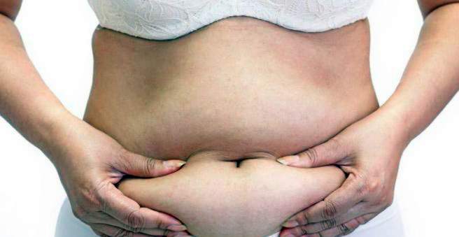
In a rectus diastase (out of alignment, midline fracture) the straight abdominal muscles diverge. This causes abdominal wall fractures. The cause is an abdominal wall weakness, which is often caused by pregnancy or obesity. The therapy consists in the strengthening of the abdominal muscles, in some cases must be operated on. Read more about symptoms, diagnostics and therapy of rectus diastase.

Rectus diastase: description
As a rectus diastase, the doctor refers to the expansion of the so-called Linea alba, a vertical connective tissue suture on the abdomen. As a result, the right and left straight abdominal muscles deviate to the side and a tactile gap remains. The linea alba is usually one to two centimeters wide and is formed by the intertwining connective tissue structures of the straight abdominal muscles, which obscure the surface of the anterior abdomen. Rectal diastasis does not count as a true fracture, even if it is similar to a fracture on standing or under increased pressure in the abdomen due to protrusion.
The rectus diastase is most pronounced in the navel and is between one and ten centimeters long. Sometimes it extends from the costal arch to the pubic bone.
In women, the rectus diastase is much more common than in men: The strains of pregnancy, the straight abdominal muscles are overstretched and diverged (Out of Alignement). If the abdominal muscles are weak, the muscle strands can even diverge more than a hand’s breadth. This affects the maintenance, support and support function of the muscles. Despite weight loss remains in this case, after pregnancy, an ugly protrusion back to the front abdominal wall.
In men, the rectal diastasis usually occurs limited to the area above the navel.
Abdominal muscles build up
Under the skin of the abdominal wall is the more or less pronounced layer of fat, which is separated from the musculature by a connective tissue structure (fascia). The abdominal muscles are the basis of the abdominal wall, which is still separated from the intestines by the peritoneum. The muscle layers are made up of different muscles. The flat straight abdominal muscle, for example, connects the ribcage and the pelvis. The lateral abdominal muscles envelop the straight abdominal muscles with their flat tendons on each side, where they form the so-called rectus sheath. In its center is the linea alba, which is affected by rectal diastasis.
Rectus diastase: symptoms
A rectus diastase usually causes no complaints. For those affected, a gap in the middle of the stomach is palpable. Tension can cause a visible and palpable bulge.
As pregnancy progresses, rectal diastasis can be felt with low back pain, buttocks, and hips during exercise. In particular, women who have already had several pregnancies suffer as the muscles have been repeatedly stretched.
Excess tissue and skin may protrude from the front of the abdomen, while in the last trimester of pregnancy the upper part of the uterus protrudes from the abdominal wall. With very large rectus diastases, sometimes even the outlines of the unborn baby can be seen.
The birth can be complicated by the rectus diastase, since the abdominal muscles can not be sufficiently strong used to push out the child. An upright posture and the back muscles can compensate for this.
Rectus diastasis: causes and risk factors
Most rectal diastases are acquired, especially pregnant women suffering from it, infrequently innate risk factors play a role.
Acquired rectus diastase
Pregnancy is a typical trigger of a rectus diastase. During pregnancy, the abdominal muscles stretch through the growing child in the uterus and lose their tension. In addition, the pregnancy hormone Relaxin has a relaxing effect and promotes stretching of the Linea Alba. The rectus diastase often arises in the last trimester of pregnancy, when the stomach becomes more and more expansive. Women should therefore try not to put extra strain on their tummy, for example by lifting heavy things.
Repeated pregnancies or multiple pregnancies increase the risk of rectal diastasis.
Obesity also leads in some cases to a rectus diastase, since the abdominal wall can also be overstretched by the abdominal fat.
Congenital rectus diastase
Rarely does the rectus diastase have congenital causes. In such a case, the abdominal muscles do not run parallel, but diverge upwards. The linea alba widens, causing the abdominal wall to bulge.
Rectus diastase in newborns
Rectal diastasis can also occur in newborns and infants, as the distance between the two straight abdominal muscles is comparatively wide. However, the rectal diastasis disappears as soon as the children start to walk. An operation is usually not necessary.
Rectus diastase: examinations and diagnosis
In case of suspected rectal diastasis, the gynecologist or general practitioner is usually the first point of contact. To collect the medical history (anamnesis), the doctor will first have a detailed conversation with the patient, in which he asks, for example, if someone already has several children.
Physical examination
In the case of rectus diastasis, the doctor makes the diagnosis relatively easily on the basis of a palpation finding. For this, the patient lies on his back and is asked to tense the abdominal wall, for example by raising his head. The doctor can feel well with his fingers above the navel the gap in the abdominal wall between the strained muscle strands.
When the patient straightens up, laughs or coughs, the rectus diastasis bulges out as a “ridge” between the two standing straight abdominal muscles. In women with a multiple pregnancy or a pathological increase in the amount of amniotic fluid (polyhydramnios) is often a clearly drawn abdominal muscles palpable.
An ultrasound examination is rarely necessary with a rectus diastase, it makes visible how far the rectus diastase has progressed.
Rectus diastase: treatment
To correct a rectus diastase, a training of the abdominal muscles are first used. If necessary, obesity is reduced. If there are hardly any complaints, surgery rarely takes place.
In addition, there are the following in everyday life: As long as the doctor can feel a rectus diastase, the straight abdominal muscles should not be charged or trained so that the rectus diastase does not increase. The best thing to do is to just lie across the side from lying down. At first you roll yourself completely to the side and then support yourself laterally with your arm in order to come to a sitting position.
Physiotherapeutic exercises
To strengthen the abdominal muscles and to reduce the gap there are various rectus diastase exercises. They should be completed with an experienced physiotherapist or midwife.
Angela Hellers’ rectus diastase exercises are a good way to rejoin the abdominal muscles and are recommended for patients over two centimeters in length with a rectus diastase: tensing the muscles diagonally from the hips to the shoulders through even muscle work against the therapist’s hand , While the midwife or physiotherapist holds the two straight abdominal muscles together, pull the shoulders up slightly and push slightly forward against the resistance. After one or two breaths, relax again.
Just two days after spontaneous delivery or two weeks after a cesarean section, each side can be trained once, or at most twice. If the exercise is repeated on several days, the rectus diastase returns and is usually only one centimeter wide. Even a rectus diastase that has been present for several years can be positively influenced by this exercise.
Diastasis recti surgery
As a rule, the rectus diastase does not need to be operated on. However, if the symptoms of rectus diastasis increase and there are complications in the midline and umbilical region, surgery is recommended. In doing so, the surgeon applies inner sutures, which fix the abdominal muscles in the correct position. With plastic nets, the abdominal wall can be additionally stabilized.
After the rectus diastase surgery, the patient wears an abdominal belt that compresses the abdomen and a special compression wash for about six weeks. Heavy physical activity and sports are taboo for at least four weeks.
Rectus diastase: disease course and prognosis
In most cases, the rectus diastase returns with the appropriate training. A narrow rectus diastase of only one or more centimeters in length can heal itself, after a pregnancy, the recovery process may take. If there is pain, surgery may be necessary. Complications only occur when getting out of the diastasis recti It forms a rupture and organs or parts of organs are trapped, but this is only rarely the case.