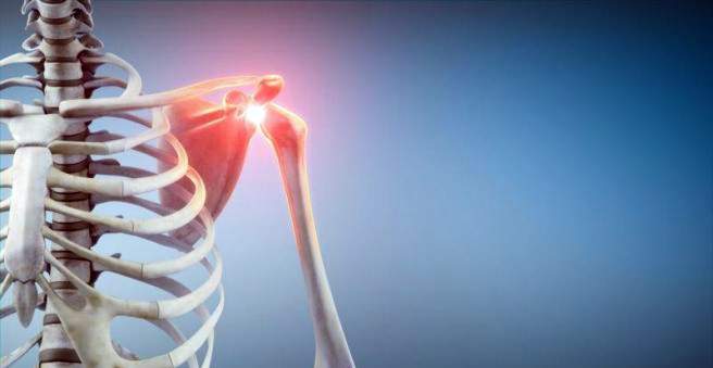
A humeral head fracture (humeral head fracture, subcapital humeral fracture) is a fracture of the head of the humerus. This type of bone fracture is most common in older people with osteoporosis, most often caused by indirect trauma. A humeral head fracture is painful and restricts the mobility of the arm. Depending on fracture type can be treated conservatively or surgically. Find out more about the humeral head fracture here.

Humeral head fracture: description
The humerus has a relatively large head, which is three times larger than the pan. This allows the shoulder a wide range of movement: The shoulder joint is the most flexible joint of the human body.
Structure of the humerus
The head (caput humeri) of the humerus is delimited from the rest of the bone by a short annular neck (collum anatomicum). This is followed by two bony prominences that serve numerous attachment points. The upper elevation is located on the outside of the humerus and is called “Tuberculum majus”. The smaller elevation is called “Tuberculum minus”.
Just below the Tuberculum minus is a thinner neck (Collum chirurgicum). Here the bone is very soft and narrow. In case of external violence, this site can break particularly easily. To the Collum chirurgicum joins the humeral stem.
humeral fractures
The humeral head fracture is counted as proximal (“proximal”) humeral fractures. The upper arm can also break in other places. When the bone breaks in the middle, it’s called a humeral shaft fracture. If it breaks at the lower end of the humerus, it is a distal humeral fracture.
Upper arm fractures near the shoulder joint account for five percent of all fractures. This makes the upper arm the third most frequent fracture site on the human body. In old age, this fracture is common, while adolescents for such a break a significant trauma is necessary.
Humeral head fracture: classification
In a humeral head fracture, the head can break into various fragments. The Oberarmkopffraktur is subdivided into four main fragments according to the doctor Neer:
- dome
- Tuberculum majus
- Tuberculum minus
- shaft
Depending on the break, two to four fragments may be created. A fragment is shifted more than an inch or twisted more than 45 degrees.
Humeral head fracture: symptoms
If there is severe pain in the shoulder area after an accident, this may be an indication of a humeral head fracture. Another sign of the fracture is the inability to move the arm or the shoulder. The area is usually swollen and tender. Furthermore, the result is an extensive hematoma, ie a bruise. He can sag after one to two days to the elbow and provide appropriate skin discoloration. In some cases, a malposition of the upper arm is visible in a humeral fracture.
Humeral head fracture: causes and risk factors
The cause of a humeral head fracture is usually an indirect trauma caused by a fall on the outstretched hand or elbow or directly on the shoulder. Osteoporosis (bone loss) plays an increasing role in older people. Due to age-related hormonal changes, bone loses firmness, becomes porous, and breaks easily. Already harmless falls can then lead to a break, such as a humeral head fracture. About 70 percent of all patients with humeral head fracture are older than 60 years.
In young people, a humeral head fracture is less common than in older and often the result of serious traffic or sports accidents (traumatic trauma). Babies may develop a humeral fracture during childbirth.
Humeral head fracture: necrosis
The more severe the injury, the higher the risk of neuropathic necrosis. The bone tissue dies from the humeral head. In a humeral head fracture with additional dislocation (luxation), the risk of necrosis is even 90 percent.
The reason for the humeral head necrosis is that the bone is no longer sufficiently supplied with blood. This occurs when certain blood vessels are injured: the Arteria circumflexa humeri anterior and its endast the Arteria arcuata as well as the Arteria circumflexa humeri posterior. The humeral head necrosis is one of the aseptic, that is not infection-related bone necrosis.
Humeral head fracture: examinations and diagnosis
If you suspect a humeral head fracture, you should consult a doctor for orthopedics and traumatology. He will first ask you about the accident and your medical history and then examine it. Some questions from the doctor might be:
- Did you fall on your shoulder or outstretched arm?
- Describe the exact accident.
- Can you still move the shoulder or the arm?
- Do you have pain?
- Did you have any complaints such as pain, restricted movement or a previous dislocation in the shoulder or arm area?
A humeral head fracture can often be identified by the nature of the accident and its symptoms. Often, the patient supports the injured arm on the wrist (as opposed to a shaft fracture of the upper arm).
Similar symptoms as a humeral head fracture show a shoulder dislocation (shoulder luxation). Therefore, the doctor will examine you for any possible nerve and vascular injuries.
Children suffering from a birth traumatic humeral head fracture often take a restraint. This is sometimes misinterpreted as plexus palsy (paralysis). This can be checked by a movement test: In a humeral fracture, the child – in contrast to a paralysis – pain when the arm is moved.
Humeral head fracture: Imaging procedures
To confirm the suspected diagnosis of humeral head fracture, X-rays are usually taken from all sides of the shoulder. The images also reveal whether fracture parts have shifted or whether other bony structures have broken.
If the fracture is only slightly shifted, it is checked whether the head fragments are stable even when the arm is gently spread out to 80 degrees laterally. Even more accurate is computed tomography (CT), which shows the exact relationship of each fragment. The CT scan is especially indicated when planning an operation.
In special cases magnetic resonance imaging (MRI) can be used to exclude soft tissue damage such as tendon injuries.
Furthermore, angiography (vascular radiography) is used to locate the site of a possible vascular injury. An electromyography (EMG) can determine if the arm vessels are still intact.
Humeral head fracture: treatment
Depending on the severity of the humeral head fracture, there are various treatment measures. In acute humeral head fracture, it is especially important to treat the pain and to avoid further damage. If you suspect a shoulder dislocation, do not attempt to retract the joint as this may cause more damage. First of all, imaging must confirm the dislocation!
Humeral head fracture: conservative therapy
In case of an uncomplicated upper arm fracture, an operation can be avoided in many cases. Unless the fragments are shifted from one another, the upper arm is usually immobilized with a special dressing (Desault or Gilchrist dressing). Accompanying a physical cold therapy can be applied (cryotherapy).
As a result, the person concerned can start with light exercises, which, however, should not be exercised in the pain area. As soon as the pain subsides, the physiotherapy begins with pendulum movements of the arm. After two to three weeks, the patient is allowed to actively and passively move the arm again.
It is important that the healing progress is observed with X-ray controls. As a rule, follow-up is after one day, ten days and six weeks. The bone is stable again after about six weeks with adequate healing.
Only rarely are displaced fractures treated conservatively. This is the case, for example, when there is a high risk of surgery. The arm is placed after the application of special associations additionally with a cast. Especially in children, a humeral fracture can often be treated well conservative, because he aligns spontaneously again.
Humeral head fracture: operative therapy
In general, there are two different surgical procedures, depending on the location and type of injury: osteosynthesis and joint replacement. The surgeon also determines, depending on the type of fracture, whether an open or a closed operation is indicated.
A humeral head fracture is basically an urgent but not an emergency operation. It should first be immobilized in the Gilchrist or Desault Bandage and operated on within ten days.
If there are concomitant vascular or nerve injuries, as is often the case with a fracture in the area of the collum anatomicum or a dislocation that can no longer be restricted, surgery is usually performed immediately to avoid lasting damage.
In a Tuberculum majus fracture, in which the fragments are displaced, the shoulder joint is often additionally dislocated. The bone is then stabilized after correction with plates, screws or drill wires. Then the arm is immobilized in a special bandage. If the fracture is not postponed, active movement of the muscles is only recommended after three weeks because of the risk of displacement.
If it is an unstable humeral head fracture with a severely displaced fracture as well as a dislocation fracture, surgery is also performed. The goal is to anatomically rebuild the head of the humeral head so that aftertreatment is not necessary.
Older people of lower bone quality who are at high risk for bone necrosis are first treated with a prosthesis. New angle stable implants show good results. In younger patients, attempts are made to maintain the humeral head and anatomically align the fracture parts.
It is recommended that the shoulder joint not be completely quiet for more than two to three weeks, otherwise a so-called “frozen shoulder” may develop – a painful stiffening of the shoulder.
Humeral head fracture: disease course and prognosis
When a humeral head fracture is operated, complications such as wound healing disorders, infections, or rebleeding rarely occur. Occasionally a humeral head fracture does not heal completely (pseudarthrosis). The function is hardly affected by this. Especially in children, the prognosis of a humeral head fracture is good.
Other possible complications of humeral head fracture include:
- Humeral head necrosis (especially in elderly patients)
- Impingement: painful pinching of soft tissue in the joint space (between the shoulder roof and the humeral head) in the case of the greater tuberculum fracture
- Labrum lesion (injury to the gel cliff)
- Rotator cuff tear (tear of the muscle group in the area of the shoulder)
- Vascular and nerve damage (such as the axillary nerve or the axillary artery) in severe humeral head fracture
The aim of the treatment is always that the upper arm in everyday life is able to move without pain. In some cases, however, the shoulder can be after one Humeruskopffraktur do not move as before. The arm can not then be moved forward and sideways to the vertical. This happens in about 10 to 20 percent of cases.