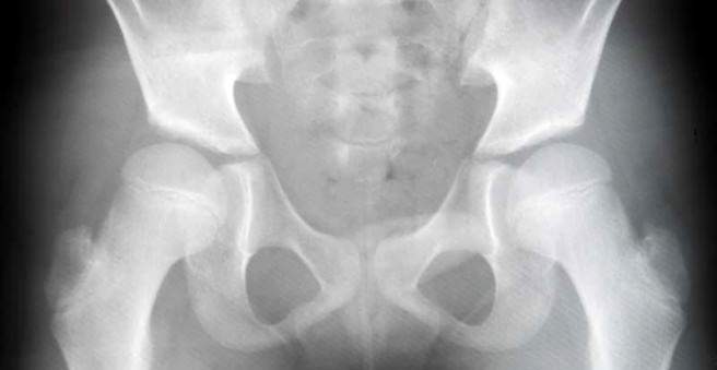
As hip dysplasia, doctors refer to a congenital or acquired malformation of the acetabulum. It occurs in about two to three out of 100 newborns, especially in girls. Left untreated, hip dysplasia can result in permanent damage to the femoral head or socket. A later disability and premature signs of wear are the possible consequences. Read all important information about hip dysplasia here.

Hip dysplasia: description
Hip dysplasia is a congenital or acquired malformation of the acetabular cup. As a result, the still cartilaginous-soft femoral head of the femur does not find a stable hold in the acetabular cup. In the most severe case of hip dysplasia, hip dislocation, the head of the femur slips out of the socket.
Hip dysplasia and hip dislocation can only occur on one hip joint or on both joints. In a one-sided malformation, the right hip joint is much more affected than the left hip.
Hip dysplasia: frequency
Of 100 newborns, two to three have hip dysplasia. Hip dislocation is much rarer with a frequency of about 0.2 percent. Girls are more often affected than boys.
Hip dysplasia: adults
Unrecognized or late-treated hip dysplasia in babies severely restricts mobility later in life and can cause pain in adolescents. It can lead to premature wear-related changes, which can limit the choice of career and may lead to early disability. Malformations of the hip joint, such as hip dysplasia, promote early joint wear (osteoarthritis).
Hip dysplasia: symptoms
Hip dysplasia alone does not initially cause any discomfort. However, if it is not detected in time, damage to the acetabulum and head (such as a hip osteoarthritis in later life) or dislocation of the hip can occur.
In a hip dislocation of the femoral head (ie the head of the femur) jumps out of the socket. In this case, the baby can spread the legs only incomplete. The leg on the affected side appears shorter than the other. The anal furrow and pubic fold are shifted to the affected side. However, leg shortening and wrinkle asymmetry may be absent in bilateral hip dislocation.
As a result of hip dislocation, the “empty” socket can gradually deform. In some cases, the head of the femur can then no longer be restricted to the normal position.
In older children, hip dysplasia may result in a hollow back or “waddling”. Such signs should prompt parents and their child to see a pediatrician or orthopedist.
Hip Dysplasia: Causes and Risk Factors
The exact causes of hip dysplasia are unknown. But there are risk factors that favor the development of this malformation:
- Incorrect position of the fetus in the womb: Children born in the lumbago or breech have approximately 25 times more often hip dysplasia than babies born in the normal birth position.
- Tight conditions in the womb, such as in a multiple pregnancy
- Hormonal factors: The pregnancy hormone progesterone, which loosens up the maternal pelvic ring in preparation for childbirth, probably causes a greater relaxation of the hip capsule in female fetuses – hip dysplasia can develop.
- Genetic predisposition: Already other family members had hip dysplasia.
- Malformations in the area of the spine, legs and feet
- Neurological or muscular diseases such as open back (spina bifida)
- Malposition of the hip joints after birth
Hip dysplasia: examinations and diagnosis
As part of the screening examinations, the pediatrician routinely checks every child for hip dysplasia already at U2 (third to tenth day of life). For a reliable diagnosis, he then performs an ultrasound examination of the hip at the U3 (in the 4th to 6th week of life). X-ray examination to diagnose hip dysplasia is usually unnecessary and also less reliable, as the still cartilaginous baby bones are less recognizable in the X-ray than in the ultrasound.
On physical examination, the following signs may indicate hip dysplasia:
- Glutealfold asymmetry (unevenly formed skin folds on thigh base)
- Abspreizhemmung (a leg can not be spread as far as usual)
- Unstable hip joint
Hip dysplasia: treatment
The treatment of hip dysplasia depends on the severity of the changes. Both conservative and operational measures are available.
Conservative treatment
The conservative treatment of hip dysplasia or hip luxation consists of three pillars: maturation treatment, reduction and retention.
Ausreifungsbehandlung:
A hip instability at birth due to a delay in maturity is inherited in 80 percent of normal motor development cases within two months. As a medical measure usually sufficient monitoring by means of ultrasound. The maturation can be supported by wrapping the child with extra-wide diapers.
In a higher-grade hip dysplasia, but in which the femoral head is still in the joint socket, the baby is a Spreizhose or Abspreizschiene adapted. The duration of treatment depends on the severity of the dysplasia and continues until the formation of a normal acetabular cup. This process is checked at regular intervals by means of ultrasound. In rare cases, as soon as the acetabular cup has matured at twelve months of age, the doctor will make an X-ray of the hip. He can check whether the femoral head and cup are well formed.
Reduction and retention:
If the femoral head of a child with hip dysplasia has slipped out of the socket (dislocation), it must be “restrained” in the acetabulum (reposition) and then held there (stabilized) (retention). For children less than nine months old, a repositional bandage may be used, in which the hip joints can spontaneously contract due to the child’s kicking and are then stabilized in this position for a longer time by the bandage.
Another possibility is the manual Einreken the “slipped” femoral head and then applying a plaster in seat squat position for several weeks. He keeps the femoral head stably and permanently in the acetabular cup. The restored contact allows the head and socket to develop normally.
If the hamstring did not work or if the affected child is already older, an extension treatment is often carried out in preparation. It serves to loosen the hip joint and stretch the shortened muscles.
surgery
If conservative measures for the treatment of hip dysplasia are unsuccessful or the malposition is detected too late (in children who are three years or older, or in adolescents or adults), surgery is necessary. There are various operational procedures available for this purpose.
Hip Dysplasia: Prevention
Hip dysplasia can not be prevented. However, wide wrapping causes babies and toddlers to spread their legs more. This is considered beneficial for the hip joints.
For the complete cure of hip dysplasia, it is crucial if it was detected early. Therefore, a doctor should examine babies for U2 screening, but no later than U3 for hip dysplasia. Early therapy reduces the risk of permanently damaging the femoral head or the acetabular cup.
Hip dysplasia: disease course and prognosis
The earlier hip dysplasia is treated, the faster it can be repaired and the greater the chances of recovery. With consistent treatment in the first weeks of life and months of life, develop the hip joints in more than 90 percent of children affected normal. However, if hip dysplasia is detected late, surgery is usually unavoidable. In addition, a hip dislocation and premature wear of the hip joint threaten – an arthrosis even in young adulthood may be the result.
Among the risks of surgery and reduction include stunting of growth of the femoral neck and a so-called Hüftkopfnekrose, that is, a death of the femoral head.
However, if a hip dysplasia is not treated, it will lead to a malformation of the joint socket and in the future to a disability.
At a hip dysplasia helps physiotherapy to counteract a limp. In particular, those muscles are trained, which stabilize the hip.