Pressure ulcer is the medical term for a pressure sore. Colloquially, one also speaks of bedsores. The cause is permanent, heavy pressure that can damage the skin and underlying tissue down to the bone. Bedridden people and wheelchair users are particularly prone to pressure ulcers. With a careful prophylaxis, the pressure ulcers can be avoided. Read all important information about the development, treatment, prophylaxis, diagnosis and course of pressure ulcers.

Quick Overview
- What is pressure ulcer? Pressure ulcer, which occurs especially in places where bones are close to the surface of the skin (buttocks, elbows, heels, ankles, etc.). Above all, those affected are largely unable to move, bedridden patients and wheelchair users.
- Reason: Persistent, strong pressure that squeezes the blood vessels. Affected tissue is poorly supplied with blood, acid metabolites are no longer transported away and gradually destroy skin, tissue and bones.
- Risk factors: Long, motionless sitting or lying, thin, less elastic skin, diabetes, reduced sensitivity to pain, low body fat, incontinence, certain medications, obesity, lack of care, malnutrition, existing skin diseases and irritations.
- Treatment: moist wound dressings and regular cleansing. Removal of dead tissue. If the pressure is high, surgery may be necessary. It is also important to eliminate the cause of the pressure ulcers, such as pressure-relieving aids (anti-decubitus mattresses or seat cushions), regular repositioning, etc.
- Possible complications: The wound can infect and then cause complications such as bone marrow and bone inflammation, pneumonia, bone abscesses or blood poisoning.
- Prophylaxis: Anti-decubitus aids (foam mattresses, gel or air cushions, sheepskin pads, special seat cushions for wheelchair users, etc.), regular transfer and mobilization of bedridden patients, frequent changes of clothing and bedding, skin care, balanced nutrition, adequate hydration etc.
- History and prognosis: Lengthy healing process even with optimal treatment. Increased risk of relapse after successfully treated bedsores. Therefore, physicians recommend a careful prophylaxis and the earliest possible treatment of pressure ulcers.
Pressure ulcers: description
A pressure ulcer (decubitus, decubitus ulcer) is a localized damage to the skin, the underlying tissue and in extreme cases, the bone. It shows up in the form of a different deep, permanently open wound (eg on the buttocks, tailbone or on the heels). Especially bedridden people are affected. Experts estimate that about every tenth to thirtieth patient in the hospital develops a pressure sore. In nursing homes, it is even 45 percent. Even wheelchair users have an increased pressure ulcer risk, especially in the buttocks area.
Pressure ulcers are very painful, In addition, they can infect, A first indication is one unpleasant foul odor from the wound, On the psychological well-being, the pressure sores can affect. Because they restrict the patients in their daily lives. The persistent, painful wounds can even lead to depression.
Pressure ulcers: graduation
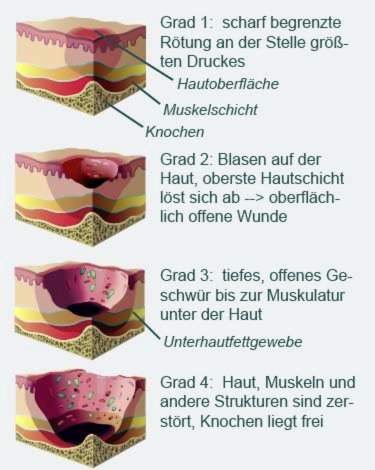
A pressure ulcer changes the skin. Depending on how strong the changes are, there are different degrees of severity:
- Decubitus grade 1 (stage I): In the initial phase, the affected skin reddened and sharply separated from their surroundings. The redness persists even when the pressure subsides. The area may be hardened and warmer than the surrounding skin, but the skin is still intact.
- Decubitus grade 2 (stage II): In decubitus grade 2, blisters have formed on the skin. Sometimes the uppermost layer of skin is already detached. The result is an open wound, which is still superficial.
- Decubitus grade 3 (stage III): In pressure ulcer grade 3, the pressure ulcer extends to the muscles under the skin. You can see a deep, open ulcer. Beneath the healthy skin in the marginal area of the decubitus are sometimes “pockets” that emanate from the ulcer.
- Pressure ulcer grade 4 (stage IV): In a stage IV pressure ulcer, one looks at exposed bones. Skin, muscles, bones and other structures such as joints or tendons are destroyed.

Where decubitus is particularly easy to form
Some parts of the body are particularly sensitive to pressure, so that decubitus can develop quickly there. Endangered are areas where bony prominences are directly under the skin, without being protected by fat or muscle tissue. Examples of this are buttocks,the big rolling hills (trochanters) on the outside of the thighs,ankleandHeels.
In the supine position, decubitus most commonly occurs on the buttocks, over the coccyx and on the heels. In lateral position are usually affected the rolling hills of the thighs and ankles. Rarely, decubitus develops on the ears, back of the head, shoulder blades or toes.
Basically, pressure ulcers form less frequently in lateral or prone position. An exception is prolonged prone surgery, which can cause pressure sores on knees, on the face (forehead and chin), on the toes or in the pubic area.
Pressure ulcers: complications
If a pressure sore is not treated quickly, it spreads to deeper tissue layers. The tissue dies (Necrosis) and must be surgically removed. In addition, the wound can be infect, If the infected ulcer is already in the bone, the pathogens can also spread there. It can become one Bone inflammation (Ostitis) and Bone marrow inflammation (osteomyelitis) develop. If the germs spread further in the body, this can be one Pneumonia, bone abscesses or one Blood poisoning (sepsis) trigger.
Also deficiency These can be the result of decubitus if it spreads extensively on the skin. Because then the affected person permanently loses important minerals and proteins via the open wound.
Pressure ulcers: development
A pressure sore develops when the tissue becomes permanently attached strong pressure is exposed. Then the small blood vessels are squeezed and the cells are no longer sufficiently supplied with oxygen and nutrients, so that they die.
In addition, the blood can no longer flow through the veins. This will accumulate acid metabolites in the tissue. In healthy people, the resulting pressure pain triggers a knee-jerk movement that relieves pressure from the affected area of the body. The tissue is then perfused again better.
Unlike older people and patients with one disturbed sense of pain: With them, this movement reflex is often weakened. So the pressure remains and the tissue is acidified. As a result, the arterial blood vessels are widely deployed to more effectively bleed the tissue. You can tell by the fact that the skin is reddening. The dilated vessels deliver fluid and proteins to the adjacent tissue, resulting in Water retention (edema) and Blow form. The tissue is increasingly destroyed – a decubitus has developed.
Pressure ulcers: risk factors
Various factors favor the development of pressure ulcers:
- Long lying or sitting: Pressure ulcers develop especially in people who lie or sit motionless for a longer period more or less motionless. Often the pressure ulcers occur in elderly patients who are bedridden due to an acute or chronic disease. Even patients in wheelchairs belong to the risk group.
- Thin, inelastic skin: The skin of older people is often dry, limp and inelastic. Thus, it is particularly sensitive to shear forces (shifts of tissue layers against each other). If patients slip restlessly around the bed, the resulting friction can severely damage the thin skin and promote the development of a pressure sore.
- Diabetes (Diabetes): Diabetics are particularly susceptible to pressure ulcers: Diabetes causes damage to nerves over time, so that diabetics can no longer feel their touch, pressure and pain. Delayed accordingly, they register increased pressure on skin and tissue.
- reduced sensitivity to pain
- low body fat percentage
- incontinence: Leads to moist skin on anus or vagina. The skin softens, which promotes pressure sores.
- certain medications, for example, pain medication
- overweight: Increases the pressure on skin and tissue when lying down or sitting.
- lack of care: Long lying in unaltered diapers softens the skin, leads to irritation and favors a decubitus ulcer.
- Malnutrition / malnutrition: It dries out the skin. In addition, those affected lack pressure-cushioning fat deposits. Both pressure ulcers pave the way.
- existing skin diseases and irritations
Pressure ulcers: treatment
The earlier a decubitus is detected, the better it can be treated. Basically, the therapy is divided into two areas: local and causal therapy.
local therapy
The local therapy aims to supply the pressure ulcer and aid its healing. In the case of first-degree decubitus, it is usually sufficient to care for the affected area of the skin carefully and to relieve pressure.
Pressure ulcers in advanced stages must be freed from dead tissue (debridement). This is done either surgically with the scalpel (surgical debridement), using enzymes (enzymatic debridement) or fly larvae (biosurgical debridement, madness therapy). The wound is then disinfected, covered with moist wound dressings and cleaned regularly.
Sometimes, as part of the local therapy and technical procedures such as Vacuum sealing method for use.
causal therapy
A pressure ulcer can only be treated successfully if its cause is eliminated: the pressure. For lying patients, for example, a special one is recommended Decubitus mattress or a special bed, In addition one should the patients relocate regularly, Wheelchair users are seat cushions advisable. Read more about this later in paragraph Pressure Ulcer: Prophylaxis.
The right nutrition also plays an important role: A diet rich in protein, vitamins and minerals helps the skin to recover better and prevents malnutrition. Sometimes one will specialty foods administered.
Help against the pain associated with pressure sores Painkiller, In addition, special promote exercises the blood circulation and prevent the patient always lying in the same place.
Also part of the causal therapy is the effective Treatment of comorbidities, also of a psychic nature. For example, depression can threaten treatment.
Pressure ulcers: surgery
Pressure ulcers grade 1 to 3 (stages I to III) usually do not have surgically removed become. Differently, however, with decubitus grade 4 (stage IV): Here almost always a surgical intervention is necessary. The surgeon cuts out the pressure sore. Sometimes a part of the bone has to be removed.
For very large pressure sores, a plastic surgery to be necessary. The surgeon then transplants skin and soft tissue from other parts of the body to the damaged part of the body.
Photos can help to understand the success of the treatment. So do not be surprised if the medical staff regularly photographs the wound.
Pressure ulcers: prophylaxis
Physicians and nurses regularly value each individual patient Decubitus Risk from. For this purpose, documentation sheets are used, for example the so-called Braden Scale, For certain risk factors, such as the degree of physical activity of the patient, his mobility and his ability to respond to pressure-related complaints, between one and four points are awarded.
At the end of the scale all points are added. A score of 18 points or more means no pressure ulcer risk. The lower the number, the higher the risk. If the score is less than nine, the patient has a very high risk of pressure ulcer.
Based on the result individual measures for decubitus prophylaxis met. Examples:
Anti-decubitus aids
Anti-decubitus aids reduce the pressure on vulnerable areas of the skin by spreading it more evenly. These systems have proven themselves:
- Soft storage systems such as foam mattresses, gel pads or air cushions distribute the body weight and thus the pressure on a larger area.
- Alternating pressure systems (Alternating pressure mattresses) consist of differently arranged air cushions, which are alternately inflated with air. Some systems are equipped with software that detects when the pressure in certain places becomes too strong. The system then automatically initiates a displacement by venting the air from the respective chambers and filling other chambers.
- Micro-stimulation Systems (MiS) are a type of electrically controlled decubitus prophylaxis mattresses. They promote the patient’s own movement through their own small movements. This stimulates blood circulation in the tissue, which prevents pressure sores or supports the wound healing of existing ulcers.
Also sheepskins are well suited as a pressure-relieving pad. However, they give off a lot of heat and are therefore not always perceived as pleasant.
For wheelchairs special anti-decubitus seat cushions are suitable. These reduce the pressure on the buttocks.
Rearrangement and mobilization
Bedridden patients must be regularly transferred – even if they are lying on an anti-decubitus mattress. In addition, the patients should be mobilized using targeted movement exercises. This includes mental mobility as a central requirement for physical activity. Reading, listening to the radio, hobbies, conversations – all this stimulates the mind and keeps the body fit.
skin care
Careful skin care keeps the skin healthy and thus reduces the pressure ulcer risk. Proper skin care means with regard to decubitus prophylaxis:
- Wash the skin with the coolest possible water.
- Forgo washing additives largely or prefer liquid, wash-active substances.
- For very dry and brittle skin, use oil bath additives.
- Use personal care products such as creams and lotions adapted to the skin type of the patient (e.g., water-in-oil products for normal to dry skin; oil-in-water products for oily skin).
Right nutrition
Although it is not possible to prevent decubitus via nutrition, it reduces the risk of developing it. Experts recommend one wholesome, varied diet, For existing pressure ulcers sufficient vitamins and minerals must be supplied to promote wound healing. In addition, at-risk patients should drink a lot to prevent the skin from drying out.
Further measures
The following measures also help to reduce the pressure ulcer risk:
- Frequent changing of clothing and bedding in patients who are sweating or incontinent. This prevents the skin from softening due to the moisture.
- Use breathable incontinence garments
- Place buttons and seams of bedding and bedding so that they do not press on the vulnerable skin
- Therapy of basic and concomitant diseases (diabetes, depression, etc.)
Anyone who takes care of bedridden relatives at home can learn in special courses for decubitus prophylaxis how pressure ulcers can be effectively prevented. The course content includes information about suitable storage and relocation techniques as well as tips for suitable aids and care measures.
Pressure ulcers: examinations and diagnosis
A pressure ulcer is visible to the naked eye. That’s why people at risk should be particularly prone to pressure ulcers prone areas of the body regularly inspected become. If possible, you should show the patient how they can examine their own skin. For example, you can look at your own buttocks with the help of a mirror. Also, the partner or friends can be involved to assess the corresponding body sites regularly. In clinics and nursing homes, the nursing staff does this important job.
finger test
With the so-called finger test, a decubitus can be identified early. To do this, press your finger on the already reddened, suspicious skin area. If the skin is not noticeably paler immediately after releasing it, it is already a pressure ulcer in stage I (grade 1).
Smear, blood test, x-ray
To detect infection of the wound, the doctor usually takes a tissue sample of the affected area. It is examined for germs in the laboratory. Also, fever, chills and an unpleasant foul odor from the wound may indicate an infection. In addition, the patient’s blood can be examined for pathogens.
For a very advanced pressure ulcer, an X-ray is also advisable. This is to determine whether the pressure ulcer has already penetrated into the bone.
Pressure ulcers: disease course and prognosis
How long it takes for a patient to develop decubitus depends on many factors. In addition to the age of the patient, among other things, his nutritional status and the cause of bed restraint play a major role. Some people develop decubitus within a few hours. This can happen if the skin is damaged by other diseases or the metabolism is disturbed (such as in diabetes).
An advanced pressure ulcer heals even with optimal treatment only slowly. Sometimes it takes months for the pressure ulcer to completely disappear. That is why it is so important to pay attention to a careful decubitus prophylaxis and to react quickly in an emergency.
However, even after a pressure sore has healed, patients have an increased risk of developing decubitus again at the affected site (recurrence). Therefore, you should control the affected area of the skin very intensively and carefully protect it from pressure. This can prevent another bedsore forms.
Additional information
guidelines:
- Guideline “Cross-sectional Specific Pressure Ulcer Treatment and Prevention” of the German-Speaking Medical Society for Paraplegia e.V. (2017)
- Guideline “Prevention and Treatment of Pressure Ulcers” by the National Pressure Ulcer Advisory Panel, European Pressure Ulcer Advisory Panel and Pan Pacific Pressure Injury Alliance (2009)