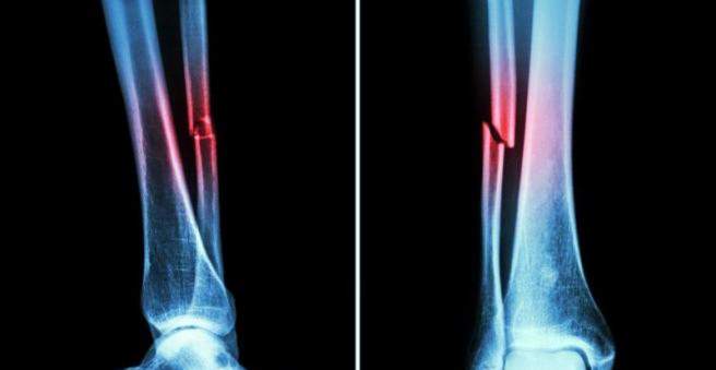
A fibula fracture and tibia fracture can be caused by direct and indirect force on the lower leg. If both bones are broken at the same time, it is also called a lower leg fracture. Swelling, pain and bruising in the affected area indicate breakage. Find out more about the fibula fracture and tibia fracture here.

Fibula fracture and tibia fracture: description
The lower leg consists of the tibias (tibia) and the fibula bone (fibula). The membrana interossea is a connective tissue membrane that connects both full length bones. A complete lower leg fracture is a joint fracture of the tibia and fibula. If one of the two bones remains intact, it is called an isolated tibial fracture (tibial fracture) or isolated fractured fibula (fibula fracture).
A tibial fracture (tibial fracture) most commonly occurs near the ankle, as the bone has the smallest diameter there. Since the lower leg is involved in both the knee and the ankle, in a lower leg fracture and the two joints may be affected. A special form of ankle fracture is the Maisonneuve fracture: a high isolated fibula fracture, in which the ligamentous structures between the tibia and fibula (syndesmosis) and the connective tissue membrane around the two bones (membrana interossea) are also injured. Often the inner ankle is broken.
The fracture of the tibia and fibula are divided into different types of fractures according to the type and location of the fracture according to the AO classification:
- Type A: only one broken line, two bone fragments
- Type B: wedge-shaped bone fracture line, three bone fragments
- Type C: rubble break with three or more bone fragments
Fibula fracture and tibia fracture: symptoms
In a splinting and fibula fracture usually complains of significant pain. It is impossible for him to burden the leg or to bend the lower leg in the knee. Other typical symptoms include swelling and bruising in the affected area. Accompanying are often grazes and soft tissue injuries.
A splint or fibula fracture may be open or closed. In an open fracture of the skin and soft tissues are injured, so that the bone fractures are visible. An open shin fracture is particularly common as the tibial leading edge is surrounded by only a small soft tissue sheath. There is always a high risk of wound infection, because bacteria can easily penetrate through the open wound.
A closed shin fracture or crush injury can cause a so-called compartment syndrome: Blood or swelling can squeeze the muscles, blood vessels and nerves in the fasciae (compartments). This causes a lot of pain. In the extreme case, the tissue can die off.
Isolated fibula fracture symptoms are rare. The fracture can often be overlooked, as the tibia is the load-bearing bone, and patients often can still walk normally despite the fractured fibula. Even with a Maisonneuve fracture, in which the fibula is far above and the inner ankle are broken, the complaints usually occur only on the ankle.
Fibula fracture and tibia fracture: causes and risk factors
A fracture of the tibia and fibula results from a direct or indirect trauma. If the lower leg is bent or twisted, indirect forces act on the leg. This can happen in a snowboard accident. If the fixed foot is pulled in the opposite direction than the rest of the body, a lower leg fracture can result.
Direct trauma usually requires a greater force. The break occurs in traffic accidents, for example, when a pedestrian is hit by a car or during sports, when, for example, a football player steps against the leg of a teammate. Often, additional soft tissue damage occurs.
An isolated fibula fracture occurs in a direct force on the outer lower leg or as Umknicktrauma.
In multiple injuries, a splint and fibula fracture often occurs as a chain injury. For example, the thigh, lower leg and foot of the same leg are broken.
Fibula fracture and tibia fracture: examinations and diagnosis
A doctor for orthopedics and trauma surgery is the right contact for the diagnosis and treatment of tibia and fibula fracture. He will first ask you about the accident and your medical history. Some questions from the doctor might be:
- What does the accident happen?
- Do you have pain?
- Can you strain the leg?
- Can you move the foot or bend the knee?
- Did you already have complaints like pain and restricted mobility?
The doctor will then examine your leg closely and also pay attention to possible accompanying injuries. When examining the lower leg, an audible and noticeable crunching (crepitation) can be a sure sign of a lower leg fracture. In addition, the physician checks the peripheral pulses, the sensitivity of the foot, and the motor function of the foot muscles.
Fibula fracture and tibia fracture: imaging
To further diagnose a fracture of the tibia and fibula, the leg is x-rayed, from the side and from the front. The images are taken to ensure that the adjacent joints are detected – possibly they are also injured. If the pulse can no longer be felt or if there is a visible circulatory disturbance, an ultrasound examination (Doppler sonography) is carried out immediately. If the examination does not yet provide a clear finding, the vessels are examined by means of angiography (vascular X-ray).
Fibula fracture and tibia fracture: treatment
Depending on the type of fracture, a fibula fracture and tibial fracture are treated conservatively or surgically.
Tibia and fibula fracture: conservative treatment
Conservative treatment, for example, usually suffices for closed, simple fractures with few bone fragments. Also, fractures in children are usually treated conservatively, if the bone parts are not shifted or the bone is incompletely broken.
Until the swelling subsided, the leg is immobilized in a split plaster. Thereafter, the gypsum can be circulated and must be worn for about two to four weeks. Thereafter, the patient gets a walking plaster for four weeks or a Sarmiento plaster, with which the knee can be bent.
The immobilization of the leg is a risk of thrombosis: it may thus form a blood clot that clogs a blood vessel. Thrombosis prophylaxis is therefore very important.
Tibia and fibula fracture: surgery
It is always operated if there is an open fracture, a fractured fracture, a fracture fracture or a break with vascular and nerve injuries.
In a shin fracture, an intramedullary nail is inserted into the marrow of the long bone to stabilize it. Doctors call this operation also intramedullary nail osteosynthesis. For more complex fractures near joints, the fracture is often stabilized with a metal plate (plate osteosynthesis). The Im
In debris or defect fractures with significant soft tissue damage, the lower leg is externally stabilized with a fixator external. This is often done in multiple injured (polytraumatized) patients.
In children, due to the growth joints, usually no nail fixation is used. The fracture is instead stabilized with a fixator external or a so-called elastic-stable intramedullary nailing.
Implanted material (such as plates, intramedullary nails) will later be surgically removed – at the earliest after 12 months.
Fibula fracture and tibia fracture: disease course and prognosis
The duration and course of the healing process are different and depend essentially on the accompanying soft tissue injuries. If the soft tissues are intact, the healing process is much better. In contrast, fractures with soft tissue injuries and defect fractures are often associated with complications.
Fibula fracture and tibia fracture: complications
A fibula fracture and fracture of the tibia can cause a number of complications. For example, vessels and nerves can also be damaged. If the bone heals with delay, a pseudarthrosis can develop. If a break does not heal in the correct position, this can lead to an axis turning error. Among the other possible complications in one broken fibula and tibia fracture include infections and wound healing disorders.