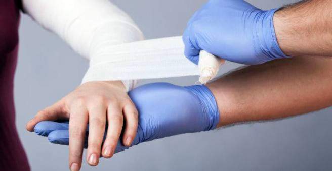
The fracture treatment depends on various factors such as the location, type and extent of the fracture as well as possible accompanying injuries. In principle, a bone fracture can be treated conservatively (for example with a plaster cast) and surgically. Everything important to the possibilities of fracture treatment and the complications that can occur, read here!

The goal of bone fracture treatment
The goal of fracture treatment is to restore the normal function of the fractured bone as soon as possible. If it is a displaced (dislocated) bone fracture, the fragments should be brought back to the original position and compensate for axis misalignments. A non-dislocated fracture can usually be treated conservatively.
During fracture treatment, the bone fracture ends are quickly aligned to the original position and adequately and consistently fixed to allow rapid fracture healing. In addition, early and functional post-treatment of the limb contributes significantly to healing.
In general, treatment is based on the three principles that can be used to accelerate bone fracture healing:
- anatomical orientation of the bone
- Immobilization and fixation
- Possibility of early functional aftercare
Basically conservative and operative procedures are available:
Conservative fracture treatment
In the conservative bone fracture treatment, the fracture ends are initially aligned properly and immobilized with a plaster splint or orthosis.
The following types of fractures are generally treated conservatively:
- Shank fracture of the arm in the growing age
- rib fractures
- stable fracture on the pelvic ring
- stable vertebral fracture without constricted spinal canal
- Fracture of the clavicle
- Fracture of the scapula without joint involvement
- Little dislocated fracture of the humerus
- Fracture in the humerus shaft area
- distal radius fracture
Conservative-functional treatment
The conservative bone fracture treatment is based on the fact that the bones stabilize themselves and the muscles can serve as a splint. If you lose the pain, you can start moving slowly. Thus, the function of the extremity can be preserved during and with the completion of the healing.
The fracture is stabilized by special bandaging techniques. Pressure is exerted on the muscles surrounding the bone, which also prevents the ends of the fracture from shortening. Special splints make the fracture quiet and allow for quick healing. Depending on the progress of healing, the limb may become increasingly strained. In the case of a fracture in the area of the shoulder girdle, this is immobilized, for example, with a backpack bandage.
Conservative-immobilizing treatment
If it is a displaced or shortened fracture, it is held in place by stretched or plaster casts. This prevents a new misalignment.
At the traction In local anesthesia, a so-called Steinmann nail is hammered in, which is connected to a stirrup and to which a different weight is suspended via a pulley. A stretch bandage prevents shortening and aligns the bones along the longitudinal axis.
One plaster cast is designed to include both adjacent joints. A plaster must be well padded, so that no tissue damage caused by too much pressure. In the case of a fresh fracture, no circular plaster may be applied due to the occurrence of swelling. This results in too much pressure on the tissue and blood circulation is impaired. This can promote the formation of blood clots (thrombosis). The best is a plaster rail. However, if a circular plaster is necessary due to the fracture, it should be split to the last thread, so that blood circulation, nerves and skin are spared.
For thrombosis prophylaxis, for example, in a leg plaster low molecular weight heparin can be injected daily. In addition, sufferers should always store the leg up and cool it with an ice pack.
If the pain increases despite gypsum conditioning, this is always an alarm sign. There is a risk that, for example, a compartment syndrome or a Volkmann contracture has formed. Tissue pressure and compression can cause permanent damage.
Operative bone fracture treatment
Surgery is an option if the bone fragments do not have sufficient contact or displaced fractures can not be properly positioned. An operation is also performed if, after conservative treatment, a malposition occurs again or the affected limb can no longer be immobilized. This is the case, for example, in old patients due to the danger of thrombosis. With surgery, the injured limb can be stressed and stabilized earlier than conservative treatment. It is especially important that joint fractures heal to prevent osteoarthritis.
During surgical treatment, the fragments are anatomically precisely positioned and fixed with plates and lag screws (osteosynthesis). This allows the bone to grow directly into the opposite cortex (cortical bone). A callus does not form, therefore one speaks of direct fracture healing.
In screw osteosynthesis, the bone fragments are fixed with screws. Depending on the place of use, there are different threads for cancellous bone (the inside of a bone) and the cortex (bone cortex). Furthermore, one distinguishes compression and lag screws.
In some cases, screws alone are not sufficient to fix a fracture. Then an extra plate osteosynthesis help: An inserted metal plate serves as a splint to absorb pressure, bending and torsional forces. The plates are differentiated according to their function: They can neutralize, compress, support, bridge and anchor with angular stability.
In a fracture of the long bones (such as femoral or tibial bone) offers an intramedullary nail osteosynthesis: Here, a nail is inserted into the medullary cavity of the bone. He shines the bone from the inside, making the fracture is relatively stable and fast loadable. In a patient with multiple injuries (polytrauma), this procedure is not recommended because bone marrow particles can enter the pulmonary circulation and cause fat embolism there.
The tension-belt osteosynthesis is used in demolition fractures such as the kneecap. Here, a wire loop is used in figure eight.
External fixation stabilizes the bone externally. Through small skin incisions long screws are screwed into the bone, which are stabilized by rods. Thus, neither soft tissue nor bone in the area of the fracture pressure is exerted. This method is especially used for open or infected fractures. The disadvantage, however, is that the fracture often can not be ideally reduced and healing is therefore usually delayed.
Dynamic screw systems are another way of surgical fracture treatment. For fractures of the femoral neck, the dynamic hip screw (DHS) is used. The fracture is splinted from the inside and compressed under load. Similarly, the femur nail (PFN) of the femur, also called gamman nail works.
In composite osteosynthesis, bone cement is added in addition to the screws or plates. This method is always used when the screws in a bad bone substance find no support. This often affects older patients with osteoporosis or tumors that have destroyed the bone.
Fracture: complications
A fracture often causes complications, as the surrounding structures are often damaged as well. The following complications can occur:
Ligament injuries: In case of joint fracture or joint near the fracture, the surrounding ligaments are usually injured.
Blood loss: The fracture can tear blood vessels in the bone, the periosteum or the musculature and form a fracture hematoma. In extreme cases, the high blood loss can cause a shock.
Skin and soft tissue damage: A dislocation fracture should immediately be aligned with the axis again, in order to relieve any squeezed soft tissue. If there is swelling, surgery should be avoided. It can be tested by folding the skin in the area with your fingers. If the skin does not fold, there is swelling.
compartment syndrome: Swelling and bruising can increase the pressure in the hardly stretchable muscle lodge (muscle lodge = group of muscles surrounded by a fascia), which, if left untreated, can lead to the death of muscle tissue. Such a compartment syndrome can in principle develop with every break. If the person complains of severe pain that has been treated in vain, a compartment syndrome may be present. The tibialis anterior lodge is most commonly affected in the lower leg.
The main symptom is the passive strain pain in the affected region. If the tibialis anterior box of the lower leg is affected, sensory disturbances in the first toe space of the foot may be an indication. Other signs include a bulging swelling of the region and tension bubbles. The risk of a compartment syndrome is particularly high in patients in shock, as the distant regions are then less well supplied with blood. Even at the slightest suspicion of a compartment syndrome, the muscle lodge should be surgically split immediately.
Vascular and nerve injuries: Vascular and nerve injuries can also accompany a fracture. If the peripheral pulses can no longer be felt, the blood circulation can be detected by means of Doppler sonography and a pulse oximeter. Destruction of vessels should be treated with emergency angiography.
Bone fracture: treatment affects the prognosis
An early, adequate Fracture treatment has a positive effect on bone fracture healing. For any suspected fracture you should therefore go to the doctor!