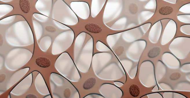
A fibroid is a benign tumor of the connective tissue. These growths are very common and can occur as a soft fibroma, hard fibroma and in many different subforms. Most fibroids are found on the skin, but also the mucous membranes may be affected (e.g., irritant fibroma on the oral mucosa). Read more about causes and therapies for fibroma.

Fibroma: description
As a fibroma, doctors call a new formation of connective tissue. This is a whole group of different growths, in which certain connective tissue cells are involved, the so-called fibrocytes. Fibromas are small tumors – but they all have in common that they are benign. Malignant tumors of the connective tissue are called fibrosarcomas.
There are different manifestations of fibroids:
Soft fibroma
The soft fibroma is also called Fibroma molle or Fibroma pendulans. These skin-colored, small tumors are common. They are common in both men and women. Especially often they are found in overweight persons. Soft fibromas often form for the first time during puberty. These are usually a few millimeters large skin protuberances. Sometimes they sit on the skin with a broad base, but often they are stalked – they hang like a small bag on a narrow approach. Colloquially, soft fibromas are also called stalk warts. They often occur on the neck, shoulders and groin. A single soft fibroma is just as possible as multiple fibroids at one site of the body; These then form a skin tumor that can grow to a few centimeters.
Hard fibroma
Most adults have at least one hard fibroma somewhere on their body. Doctors also speak of histiocytoma or dermatofibroma. They often occur on the legs, but also on the arms and on the trunk. Hard fibromas are tough nodules that are usually a few millimeters in size, rarely an inch or larger. They sit as something darker, often light brown spots in the skin. Especially often they develop in young women on the legs.
Reizfibrom
A stimulus fibroma or irritation fibroma is a fibroma of the oral mucosa. These little nodules are smooth and limited. They arise when certain parts of the mouth are repeatedly irritated.
Other fibroids
There are also some rare tumors that develop from connective tissue cells, especially in the bone area. These include:
- Ossifying fibroma: anseltener, benign tumor that occurs on the facial skull – usually in the lower jawbone.
- Non-ossifying fibroma: a pathological, connective tissue change of the bone (cortical defect), which is sometimes observed in children.
- Chondromyxoid: a tumor that usually occurs in long bones and primarily affects adolescents.
- Desmoplastic fibroma: an aggressively growing bone tumor, which occurs mainly in young people.
- angiofibroma: a tumor of the nasopharynx, which is permeated by many vessels and occurs almost exclusively in male adolescents.
The following sections are primarily concerned with fibroids affecting the skin.
Fibroma: symptoms
A fibroma is in the area of the skin visible from the outside, Soft fibroids are particularly common in the neck, armpits, groin, and women below the breasts. Often they are petiolate, and in slightly larger growths, one can see small wrinkles on the surface. Most soft fibromas are skin colored, but if you turn them, they may turn red or black due to the injury to the blood vessels.
In contrast, a hard fibroma is usually slightly darker, often grayish-brownish. They may be slightly raised or sunken relative to the skin surface. Characteristic is the so-called Fitzpatrick sign: If you push the area around a hard fibroma together with your thumb and forefinger, it sinks into the skin. This can be distinguished from a melanocytic nevus (“liver spot”).
The stimulus fibroma sits on the oral mucosa, either around the cheeks, on the side of the tongue or on the gums. It is a small, limited, smooth “Hubbel”. Its color corresponds to the surrounding tissue or is slightly lighter.
Unless they are injured, fibromas are preparing no pain.
Fibroma: causes and risk factors
The causes of a fibroma are not well known in most cases. The soft fibroma is one of the hamartomas; These are tumors that emanate from a defect in the embryonic germinal tissue. This is a tissue precursor that can later develop (differentiate) into different tissue forms. If an error occurs in the differentiation at individual points, one speaks of a Hamartie. This can result in excess tissue – in this case connective tissue – being formed. However, unlike other tumors, hamartomas do not always grow on their own. Soft fibroma is a common manifestation of a hamartoma.
There are some diseases in which people are more likely to develop hamartomas. These include, for example, the Cowden syndrome and the Neurofibromatosis type 1 (Morbus Recklinghausen). In this case, hereditary factors play a role in fibroma development. People with systemic lupus erythematosus, immunodeficiency AIDS, or a drug-suppressed immune system (such as after transplantation) often develop more dermatofibromas (hard fibroids).
When hard fibroma, experts suspect that it is made of a small Inflammation of connective tissue arises. It can have different reasons:
- insect bites
- Thorns of plants that invade the skin.
- Inflammation of a hair follicle (folliculitis)
- other minor injuries
A hard fibroma is therefore a small scar under the upper layers of the skin. Often the trigger remains undetected.
One Reizfibrom develops in places in the mouth that are often irritated, for example, by a denture or a sharp tooth edge.
Fibroma: examinations and diagnosis
For the diagnosis of a fibroma is the Dermatologists (Dermatologist) the expert. First, he asks when the changed skin spot the person affected noticed for the first time, whether it has changed or was injured. In a typical fibroma, an expert usually recognizes what it is at first glance. With a special magnifying glass (dermatoscope), the doctor can examine the fibroma in more detail. He pays attention to the size, shape, color, structure and edges of the skin change.
If it is suspected that it is a malignant growth – for example, a malignant melanoma – the doctor takes a tissue sample (biopsy). A small fibroma is usually completely removed (excision). The sample taken can be examined histologically using special procedures. So you can see how the cells are arranged and arranged.
In rare cases, so many fibromas or other lesions can be found that there is a suspicion of another underlying disease. Then follow further investigations.
Fibroma: treatment
A fibroma requires from a medical point of view no therapy, Both soft and hard fibroids are safe. There is no increased risk that they will degenerate and develop into a form of skin cancer. Normally they stop their growth from a certain size and then stay that way.
Remove fibroids
One Remove fibroma to leave is normally straightforward. The dermatologist does this in a minor procedure, depending on the size of the fibroma with a local anesthetic. Depending on the size and shape of the growth, it depends on whether the site needs to be sewn after fibroma excision.
Watch out: One fibroma In no case should you remove yourself, otherwise you risk an infection. If you have fibroma that bothers you, always visit the family doctor!
Fibroma: disease course and prognosis
Fibroma is above all an aesthetic problem. Medically, the growths of the connective tissue are completely harmless and therefore do not necessarily require a Behanldung. However, fibromas are often perceived as visually distracting, especially those on the face, neck or in the genital area. On larger fibroids, clothing and jewelry (such as necklaces) may also get caught. With a small surgical intervention, one can fibroma but usually quick and easy to remove.