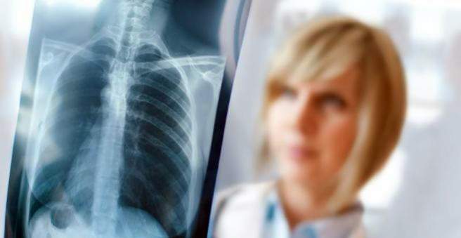
An atelectasis is understood to be airless lung tissue. Atelectasis is not an independent disease, but rather a condition that arises as a result of another underlying disease. The phenomenon can affect the entire lung, but more often circumscribed lung sections. Here you will learn how an atelectasis can develop and how it is treated.

Atelectasis: description
In atelectasis, parts of the lungs or the entire lung are depleted of air. The term comes from the Greek and means translated as “incomplete expansion”. The condition mainly affects the smallest air-carrying units of the lung, the alveoli, also known as alveoli. The alveoli have a very important function, because here the oxygen exchange takes place. The alveoli are surrounded by a fine network of tiny blood vessels, passing red blood cells absorb the oxygen from the air here. If the alveoli collapse, the affected area is no longer available for oxygen exchange. Atelectasis is therefore a serious condition.
Doctors basically distinguish two forms of atelectasis:
- Primary or congenital atelectasis: This form only affects newborns or preterm infants and is therefore also referred to as fetal atelectasis.
- Secondary or acquired atelectasis: This occurs as a result of another disease.
Atelectasis: symptoms
Atelectasis restricts lung function. The symptoms that arise as a result of this, among other things, depend on how large the affected lung section is, and whether the atelectasis occurred suddenly or gradually. The respective causes for the collapse of the lungs also shape the symptoms.
An atelectasis may occur in the affected lung section no gas exchange more to take place. As a result, the oxygen level in the blood decreases – the organism tries to compensate for this condition by accelerating the respiration and increasing the heart rate. As a sign of a lowered oxygen level in the blood, in some people affected the skin turns bluish – doctors also refer to this symptom as cyanosis.
If the atelectasis occurs quite suddenly, for example because the airways are blocked, sufferers complain of severe respiratory distress (dyspnea), in some cases also of pains in the chest. If large areas of the lung collapse, it can also lead to cardiovascular shock. The blood pressure suddenly drops sharply, and the heart beats fast (tachycardia).
An atelectasis, which is slow and affects only small parts of the lungs, causes only mild discomfort. For example, some sufferers notice that he is short of breath and quickly gets out of breath, especially when straining. It also happens that a slight extent of atelectasis is not noticed.
Symptoms of congenital atelectasis, as occurs in premature babies, often appear immediately after birth or within the first few hours of life. In the affected preemies, the skin turns bluish. You breathe fast; the areas between the ribs and over the sternum are inhaled and the nostrils move more. Frequently, affected children moan on exhalation as an expression of their shortness of breath.
Atelectasis: causes and risk factors
The innate and the acquired atelectasis can have many different reasons.
The following causes for the congenital atelectasis in question:
- immature lungs: This phenomenon particularly affects premature babies. In the alveoli there is usually a substance that reduces the surface tension of liquids, the so-called surfactant. If this substance is missing, the alveoli, which are covered by a thin film of liquid, collapse. Only in the last few weeks before birth does the lungs mature completely and produce sufficient amounts of surfactant. If a child is born too soon, however, there is a shortage of this substance and the lungs may not develop properly after birth. Physicians then speak of the respiratory distress syndrome of premature babies.
- Moved airways: If the newborn inhales mucus or amniotic fluid, the lungs can not properly fill with air. Malformations, which impede the airflow in the respiratory tract, can also lead to atelectasis.
- Disorder of the respiratory center: In the case of damage to the respiratory center in the brain (for example due to cerebral haemorrhage), the reflex may be missing air after birth.
- diaphragmatic hernia: The diaphragm (the muscle plate that separates the chest from the abdomen) is misformed and has a gap. As a result, abdominal organs can slip into the chest and squeeze the lungs so much that they have no room after birth to inflate.
Causes for one acquired atelectasis are:
- Obstruktionsatelektase: Here are the respiratory tract e.g. Clogged by a tumor, tough mucus or a foreign body.
- Kompressionsatelektase: The lung is externally, e.g. by a fluid in the chest or a very enlarged lymph node, compressed.
- Entspannungsatelektase: The cause of atelectasis here is a so-called pneumothorax. Between the skin that covers the lungs (lung skin) and the pleura, which lies against the chest wall from the inside, there is a small gap filled with fluid (pleural space). Adhesion forces both surfaces to adhere tightly to each other – thus the lungs follow the respiratory movements of the rib cage. In a pneumothorax, air enters the pleural space – the elastic lung tissue then follows its own tissue tension and the affected lung section collapses. A pneumothorax can e.g. caused by spiky injuries of the thorax or by various lung diseases.
Atelectasis: examinations and diagnosis
In most cases, typical symptoms indicate an atelectasis – in many cases, the underlying disease also suggests that there is a dysfunction of the lungs. For example, doctors in the case of premature births already expect breathing problems. Immediately after birth, the midwife and pediatrician pay special attention to the baby’s breathing, skin color, heart rate, reflexes and muscle tension. Anomalies, such as a bluish skin color or increased or too weak respiration provide clues that there are problems.
If a child is born before the 37th week of pregnancy, doctors speak of a premature birth. The immature lung of the premature baby is one of the most common causes of postpartum complications. As a rule, a pediatrician specializing in the treatment of premature babies (neonatologist) will detect atelectasis. An X-ray examination ensures the diagnosis and also indicates the degree of immature lung.
An acquired atelectasis usually focuses on the diagnosis of the underlying disease. For this, the doctor first conducts a detailed conversation with the patient and asks him about his symptoms and known illnesses. He then listens to the affected person’s lungs with a stethoscope. In the case of atelectasis, normal breathing sounds are attenuated.
If the doctor taps his chest with his fingers, the sound muffled. Here, too, the X-ray examination provides the final proof whether an atelectasis is present. Depending on the cause (for example, a lung tumor, blood or fluids in the chest, foreign bodies in the airways) further studies, such as a blood test or even a computer or a magnetic resonance tomography.
Atelectasis: treatment
The therapy for atelectasis depends primarily on its causes. The ultimate goal is to restore lung function as soon as possible and provide the body with sufficient oxygen. If, for example, a foreign body or mucous plug in the respiratory tract is the reason for the collapsed lung area, it must be removed or aspirated accordingly.
A lung tumor usually requires surgery to remove the tumor. In a pneumothorax, air has penetrated into the inter-rib gap, whereby a lung section collapses. Surgical intervention may be required in some cases, but mild forms may not always be treated.
The congenital atelectasis is usually based on insufficient lung maturity, or a lack of surfactant. To compensate for this deficiency, premature babies receive this substance as a drug. If the breathing problems are very pronounced, the baby is artificially ventilated via a thin tube in the trachea (tube).
Atelectasis: Prevention
One acquired atelectasis you can not prevent by a specific measure. Congenital forms such as respiratory distress syndrome in premature babies, however, can be counteracted to some extent. Pregnant women prone to premature birth receive a drug that promotes the unborn baby’s maturation. It is a so-called corticosteroid; Doctors usually use the active substance betamethasone here. In addition, they try to delay the birth as long as possible by means of anti-buzzing agents.
Atelectasis: Disease course and prognosis
Atelectasis is not an independent disease, but a concomitant, which can have many different causes. A general statement about the course or the prognosis is therefore not possible. Rather, the underlying disease determines the course of the disease. If this can be treated well, usually the function of the lung can be restored.
In premature babies the prognosis depends on numerous factors. Basically, the sooner a child is born, the less immature the lungs are. What problems occur in premature babies, but can not be predicted, so can extreme premature babies with a atelectasis develop well, while a later date of birth is no guarantee for a complication-free course.