Vasculitis is a vasculitis. It can affect various vessels (arteries, capillaries, veins) and cause serious organ damage. Vasculitis is caused by the body’s own defense system. Why this happens is largely unknown. Read all important information about: What is Vasculitis? Which forms of vasculitis can be distinguished? Which symptoms occur? What does vasculitis therapy look like?
Vasculitis: description
Vasculitis (plural: vasculitis) is an inflammatory disease of the blood vessels, the term distinguishes between various types of vasculitis. In all cases, proteins of the defense system are directed against the blood vessels and cause inflammation. Since the defense system is directed against one’s own body, vasculitis is a so-called autoimmune disease. And because the whole body is traversed by blood vessels, vasculitis can occur virtually anywhere.
Vasculitis-forms
Doctors distinguish a primary from a secondary vasculitis. Secondary vasculitis follows or is closely associated with another underlying disease (such as rheumatism). If one knows the exact cause, for example viral infections, physicians also speak of a secondary vasculitis.
A primary vasculitis, however, manifests itself as a separate disease. The causes of these vascular inflammations are largely unknown. Some doctors therefore also speak of idiopathic vasculitis. Frequently, these blood vessel inflammations preferentially affect very specific vessels.
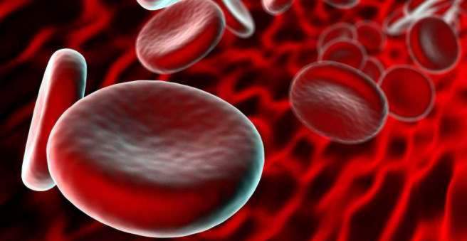
In the body there are generally arteries and veins. Arteries, also called arteries, carry the blood away from the heart. Via veins, the blood is returned to the heart. The transition between arteries and veins form the so-called capillaries (hairpin blood vessels), the smallest blood vessels of the body. They usually form a kind of vascular network through which the substance exchange takes place in the respective organ: the cells absorb nutrients and oxygen from the blood and deliver waste products to the blood.
From the heart to the target organ, the arteries get smaller and smaller. Conversely, the veins from the capillary network in the direction of the heart are getting bigger. Primary vasculitis usually affects blood vessels of a certain size. Thereafter, primary vascular inflammations are subdivided again:
|
Affected vessels |
Vasculitis-forms |
Remarks |
|
Vasculitis of large vessels |
|
|
|
Vasculitis of medium-sized vessels |
|
|
|
Vasculitis of small vessels |
|
ANCA-associated (that is, these vasculites typically have specific antipodes, the AntiNeutrophilen Cytoplasmatischen Aantibodies can be detected) |
|
Non-ANCA-associated |
giant
Everything important about this form of vasculitis you read in the article Arteritis temporalis.
Kawasaki disease
Everything important about this form of vasculitis read in the article Kawasaki syndrome.
Granulomatosis with polyangiitis
Everything important about this form of vasculitis you read in the article Wegener’s disease.
Purpura Schönlein-Enoch
Everything important about this form of vasculitis read in the article Purpura Schönlein-Enoch.
There are also endangiitis obliterans, Behçet’s disease and cerebral vasculitis:
The endangiitis obliterans (thrombangiitis obliterans) mainly affects small and medium-sized vessels of the legs. Mostly young men (<40 years), especially heavy smokers.
Behçet’s disease can be the only systemic vasculitis affecting both arteries and veins of varying sizes throughout the body. It belongs to a recent classification together with the Cogan syndrome, an autoimmune disease of the eye vessels, to “vasculitis variable vessel size”. Behçet’s disease typically occurs between the ages of 20 and 40 years. It is about three times more common in men than in women. The disease is found mainly in Turkey and in Arab countries.
Cerebral vasculitis is also called primary CNS vasculitis and affects only the vessels in the brain and spinal cord.
Vasculitis: symptoms
The symptoms differ depending on the form or location of the blood vessel inflammation. Vasculitis symptoms also depend on the extent to which the vessels are damaged by inflammation.
B symptoms
Usually a vascular inflammation begins with nonspecific symptoms. Some patients initially feel knocked off and tired. There is also a slight fever, usually below 38.5 degrees Celsius (subfebrile temperature). Some patients report heavy night sweats and unwanted weight loss.
Night sweats, weight loss and mild fever are also summarized under the term “B symptoms”. For example, cancer and tuberculosis patients also suffer from these nonspecific symptoms.
In addition to these vague symptoms of vasculitis, rheumatic complaints can also occur. Some patients complain of joint pain, which is sometimes associated with swelling. Other sufferers suffer from pain in the muscles (myalgia) and report unusually heavy muscle caters.
Vasculitis symptoms in inflammation of small vessels
The progress of vasculitis is followed by the involvement of the organs more serious complaints. These differ depending on the vasculitis form.
Inflammations of small vessels, for example, affect the eye and cause redness in the eye or blurred vision. Mucous membrane damage in the mouth leads to painful aphthae. In some vasculitis patients complain of frequent sinusitis and a stuffy nose, which sometimes bleeds. If the small vessels of the skin are inflamed, it comes to fine hemorrhages, medically called petechiae.
As a result of vasculitis of small vessels, blotchy (purpura) or bluish-tinged, net-shaped (livedo reticularis) reddening may also appear on the skin. Shortness of breath or coughing up blood may occur as part of a small vessel vasculitis of the lungs. Some patients report bloody diarrhea or bloody urine. Other forms of vasculitis cause chest pain, which may indicate myocardial or pericarditis.
If small vessels are damaged in the nervous system, sufferers report tingling or discomfort (paresthesia). These are more common on the feet than on the hands.
Vasculitis symptoms in inflammation of medium-sized vessels
In contrast, if vasculitis occurs on medium-sized blood vessels, organ infarction is the dangerous consequence. The interrupted blood supply can lead to serious damage, for example in the intestine or in the kidneys. In some cases sufferers suffer a heart attack. Inflamed vessels in the brain sometimes trigger a stroke.
Vasculitis symptoms in inflammation of large vessels
In the area of large arteries in the head, vasculitis causes severe headaches. Some of the affected patients are suddenly worse off or even completely blind. The large arm and leg vessels may close due to vasculitis and cause severe pain. Ultimately, the risk of blood clots (thrombosis) is increased. These can be carried along by the bloodstream and cause vascular occlusion elsewhere (embolism and pulmonary embolism).
Eosinophilic granulomatosis with polyangiitis: symptoms
This vasculitis is also called allergic granulomatous angiitis or Churgh-Strauss syndrome. Like Wegener’s disease, it primarily affects the respiratory tract, which usually causes asthma attacks with acute respiratory distress. In the blood white blood cells are detectable, which are also typical for an allergy.
In about half of the cases, this form of vasculitis also affects the heart. There it triggers an inflammation of the heart muscle cells or the coronary vessels. Nerves are also often damaged, and blood clots are increasingly formed which can completely occlude the vessels.
Microscopic Panarteritis (MPA): Symptoms
In this form of vasculitis, the kidneys are usually damaged (glomerulonephritis). This triggers increased blood pressure (hypertension) with headache. In case of skin involvement, small nodules and palpable bleeding under the skin (palpable purpura), especially on the legs. In some cases, nerves, paranasal sinuses or eyes may also become inflamed.
Vasculitis in essential cryoglobulinemia: symptoms
In this vasculitis variant, bleeding occurs mainly on the hands and feet. In addition, tissue defects (ulcers) may be visible. In addition, patients often report joint pain. Further vasculitis symptoms can result from kidney and nerve damage (severe course).
Cutaneous leukocytoclastic angiitis (KLA): symptoms
In this vasculitis only the small vessels of the skin are inflamed. Sufferers have skin bleeding that can be palpated and sometimes pain. In a few cases, blood-filled blisters, lumps, and non-healing ulcers may also form. KLA vasculitis most commonly occurs on the legs. Under this primary vascular inflammation women suffer about twice as often as men.
Panarteritis nodosa: symptoms
This vasculitis form affects men about three times as women. It can damage various organs, which is why the appearance of vasculitis symptoms can be very different. In most cases (80 percent), coronary arteries are inflamed. Patients often feel pressure or pain in the chest (angina pectoris) and may experience a heart attack (including younger patients). Other possible symptoms include:
- Fever, night sweats, weight loss (50 percent)
- Joint and muscle pain (65 percent)
- spasmodic abdominal pain (colic), possibly intestinal infarction (50 percent)
- testicular pain
- Stroke attack of cerebral vessels (even in young patients)
- Sensations of numbness, numbness (polyneuropathy, in about 60% of cases, mononeuritis multiplex), epileptic seizures, psychosis
- Vascular glands (aneurysms)
Up to 70 percent of the kidneys can be damaged, but not the fine renal corpuscles (no glomerulonephritis). Another important vasculitis symptom of panarteritis nodosa is the so-called livedo racemosa. It can be seen on the skin oddly shaped (lightning-like), streaky or reticulate and bluish-red skin discoloration, which does not change in appearance and remain in place.
Takayasu’s arteritis: symptoms
In this vasculitis, the main artery and its vascular branches are inflamed. It occurs mainly in Asian countries, Africa and South America. In Europe, the Takayasu vasculitis is rather rare. Women are about nine times more likely than men. In addition, the age of onset is usually less than 40 years.
Takayasu’s inflammation begins creeping (pre-occlusive stage, prepulseless phase) with mild fever, fatigue, joint and headache, and weight loss.
In the further course (Pulseless phase, occlusive stage) develop other vasculitis symptoms. Under certain circumstances, subcutaneous fatty tissue inflames and pressure-sensitive skin nodes (erythema nodosum with panniculitis) develop. Some patients complain that their arms hurt and their fingers become pale and cold (Raynaud’s syndrome).
If cerebral vessels are inflamed, visual disturbances, dizziness with fainting or a Schlaganfal can occur. Cardiac Takayasu vasculitis can lead to symptoms of coronary heart disease (CHD). This includes, for example, an uncomfortable feeling of pressure in the chest (angina pectoris).
Behçet’s disease: symptoms
If the Behçet’s disease affects the skin and mucous membranes, painful mouth ulcers (oral aphthae, 95 percent) and intimate areas (genital aphthae, 70 percent) develop. It can also form pressure-sensitive nodes (erythema nodosum).
Often, the eyes are affected, which means very often (80 percent) inflammation of the middle eye skin (uveitis). Also, an inflammation of the joints (arthritis) is not uncommon (70 percent). Almost one-third of Behcet vasculitis patients suffer from inflammation and tissue defects (ulcers) in the digestive tract. Up to 30 percent of patients also have inflamed vessels in the central nervous system (CNS). The more active the inflammations are, the higher the risk of dangerous blood clots (thromboembolisms).
Cerebral vasculitis: symptoms
This vasculitis form is also called primary or isolated angiitis of the central nervous system (PACNS). Patients often suffer from persistent dull headaches, mild dizziness and poor concentration or memory. Psychic abnormalities, such as a change in nature, also occur.
Under certain circumstances, cerebral vasculitis can sometimes no longer supply sufficient blood to brain regions, resulting in a stroke. Occasionally this is also caused by bleeding from inflamed vessels. Epileptic seizures are also associated with CNS vasculitis symptoms.
Endangiitis obliterans: symptoms
Patients often feel cold and pain at rest in their legs and feet. Superficially inflamed vessels are visible and palpable as crimson, warm and tender painful lines. Under certain circumstances, the skin appears bluish due to the reduced blood flow. In the course of vasculitis, the tissue dies, especially at the tips of the toes, and blackish skin defects become visible. In addition, nail growth may be disturbed.
Vasculitis: causes and risk factors
The causes of primary vasculitis are largely unknown. Secondary vasculitis develops as a result of another underlying disease or drug application.
Possible triggers of primary vasculitis
Primary (idiopathic) vasculitis appears as a distinct disease for no apparent reason. Some vasculitis forms, however, suspected of certain triggers.
|
Primary vasculitis |
illness |
Possible trigger |
|
Vasculitis of small vessels |
Polyangiitis with granulomatosis |
unknown, partly the bacterium Staphylococcus aureus |
|
Eosinophilic granulomatosis with polyangiitis |
unknown, questionable the asthma drug montelukast |
|
|
Microscopic Panarteriitis |
unknown |
|
|
Purpura Henoch-Schonlein |
previous respiratory tract infection |
|
|
Vasculitis in essential cryoglobulinemia |
May occur as primary vasculitis (for no apparent cause) or as secondary vasculitis (due to, for example, hepatitis C infection or malignant lymphatic system disease). |
|
|
Cutaneous leukocytoclastic angiitis |
unknown |
|
|
Vasculitis of medium-sized vessels |
Panarteritis nodosa |
unknown, association with hepatitis B infection |
|
Kawasaki disease |
unknown |
|
|
Vasculitis of large vessels |
giant |
unknown, possibly genetic predisposition and external triggering by infections |
|
Takayasu’s arteritis |
unknown |
Possible triggers of secondary vasculitis
These forms of vasculitis occur as a result of drug use, infection or other diseases.
|
Trigger of secondary vasculitis |
Examples |
|
malignant growths |
Lymphomas, breast cancer, blood cancer |
|
Rheumatic diseases |
Rheumatoid arthritis, Sjögren’s syndrome, lupus erythematosus |
|
bacterial infections |
Staphylococci, streptococci, Neisseria, Escherichia coli, mycoplasmas |
|
viral infections |
Hepatitis B virus, herpes virus, influenza virus, cytomegalovirus |
|
fungal infections |
Candida albicans |
|
Protozoa (protozoa, “protozoa”) |
Trypanosomes, Plasmodium malariae |
|
worm infections |
helminths |
|
drugs |
ASA, allopurinol, thiazides, sulfonamides, gold, NSAIDs, phenothiazines, pyrazolones, ketoconazole, tetracyclines, penicillins, propylthiouracil |
|
Food (additives) |
rare, for example, food coloring tartrazine (E 102) |
Origin of vascular inflammation
A vasculitis arises when a vessel or the vessel walls become inflamed by a defense reaction. In some of the vasculites, certain proteins of the immune system, known as auto-antibodies, are directed against the body’s own vascular structures. These vascular inflammations thus correspond to an autoimmune disease. In summary, they are referred to as pauci-immune vasculitis forms.
In contrast, there are immune vasculites. At their origin so-called immune complexes (antigen-antibody or antibody-antibody complexes) involved. Antibodies bind to certain elements of foreign substances (such as viruses, drugs), accumulate as a complex in the vessel walls and may trigger inflammation there. This occurs either at the site of complex formation (in situ immune complexes), or else the immune complexes are transported by the blood to other body regions and cause vasculitis there.
In this context, special proteins also play a role, which are usually activated by immune complexes. Specifically, these are certain glycoproteins called complement factors. They can destroy cells and cause inflammation such as vasculitis.
Consequences of vasculitis
Due to the vascular inflammation, the vessel can constrict or completely occlude. In some cases, sloughs (aneurysms) arise due to the weakness of the vessel wall. Depending on the location of the inflammation, various organs can no longer be sufficiently supplied with oxygen and nutrients. They are restricted in their function. As a result of vasculitis, in some forms, tissue nodules may form from different cells (such as epithelial cells, giant cells). These so-called non-infectious granulomas can be found in a tissue examination among others in a
- Polyangiitis with granulomatosis
- Eosinophilic granulomatosis with polyangiitis
- giant
- Takayasu’s arteritis
Vasculitis: diagnosis and examination
The specialist in diseases of the vessels is the internist. Specialists in skin and venereal diseases may be consulted in the case of vasculitis affecting the skin. If a vasculitis is suspected, the doctor first interrogates the patient and collects the medical history (anamnesis). Possible questions are:
- What complaints do you have? Do you feel choppy and tired?
- Have you lost weight inadvertently lately?
- Do you sweat a lot during the night?
- Is your temperature elevated?
- Have you noticed skin changes, such as punctate redness?
- Do you suffer from pain? If so, where and how would you describe this pain (throbbing, stabbing, etc.)?
- Did you or did you have a flu infection recently? Do you still need to cough, and possibly blood?
- Do you have any underlying conditions, such as rheumatism?
- Have you been diagnosed with infections, such as the hepatitis virus?
- Which medications do you take?
This is followed by a physical examination. Depending on the condition, the doctor will listen to and / or feel the heart, lungs and stomach. He will also measure the blood pressure on both arms and the pulse at different parts of the body. He checks the skin for changes such as unusual redness. Also, the ear, nose and throat is examined to exclude any inflammation. If a patient suffers from dizziness, fainting or skin sensation, the neurological status is also checked by means of various tests.
In vasculitis, there are usually certain areas of the body where the vessels mainly inflame. The localization of the damage already gives a first indication of what type of vasculitis it might be. To confirm the suspicion or to exclude other diseases (such as viral or bacterial infections), the doctor performs a few more tests.
laboratory tests
If the doctor suspects a vasculitis, he will take blood samples and send them to a laboratory for further investigation. As a rule, affected persons are also asked for a urine sample. Depending on the form of vasculitis, typical laboratory findings result. Inflammation levels often increase (C-reactive protein CRP, erythrocyte sedimentation, white blood cells). Of particular importance are characteristic proteins of the defense system such as auto-antibodies or immune complexes.
|
illness |
Important laboratory findings |
|
Polyangiitis with granulomatosis |
|
|
Eosinophilic granulomatosis with polyangiitis |
|
|
Microscopic Panarteriitis |
|
|
Purpura Henoch-Schonlein |
|
|
Vasculitis in essential cryoglobulinemia |
|
|
Cutaneous leukocytoclastic angiitis |
|
|
Panarteritis nodosa |
|
|
Kawasaki disease |
|
|
giant |
|
|
Takayasu’s arteritis |
|
tissue examination
To ensure the diagnosis of a certain vasculitis, the doctor takes in many cases a tissue sample (biopsy) from affected parts of the body. Samples from the skin, mucosa or kidney are taken under local anesthesia. The procedure usually takes 15 to 30 minutes.
Lung tissue is usually obtained as part of a lung (bronchoscopy). In the case of a giant cell vasculitis of the temporal artery, the physician takes a piece of this vessel at least 20 millimeters long.
The doctor sends the tissue samples obtained to a laboratory, where they are examined histologically. For example, in a vasculitis, defense cells (such as eosinophilic or neutrophilic granulocytes, antibodies and immune complexes), granulomas and other minor changes can be detected under the microscope. In this way, a kidney involvement (glomerulonephritis) can be accurately determined.
imaging
Imaging studies help to rule out other causes of the symptoms described, such as cancer, bleeding or vascular changes. For example, magnetic resonance imaging (MRI) or computed tomography (CT), and sometimes positron emission tomography (PET) are also used. In many cases, contrast agents are used during these examinations in order to be able to better assess vessels (angiography).
An X-ray image can also contribute to the diagnosis of Wegener’s vasculitis of the lungs. Here, the doctor can recognize, among other things, conspicuous round shadows in the lung tissue. Another imaging study is ultrasound (sonography), called heart echocardiography. Thus, for example, the blood flow (color duplex sonography) and vessel narrowing or -aussackungen be checked. For joint examinations the ultrasound is also suitable.
Further investigations
Especially in suspected Behçet vasculitis, the doctor performs the so-called pathergie test. To do this, he injects a small amount of saline solution into the patient’s lower arm skin. In the case of patients, an overactive skin reaction occurs within 48 hours (marked reddening, formation of a visible and palpable nodule, sometimes also pus-filled blisters). A similar reaction can also be seen in leukocytoclastic and Wegener’s vasculitis.
If the patient shows vasculitis symptoms of the brain or spinal cord, the physician will take a sample of the cerebrospinal fluid (CSF) for further analysis in the laboratory.
Many doctors necessary for the vasculitis diagnosis
In some cases, the patient has to introduce himself to various doctors in order to rule out individual illnesses and confirm the vasculitis. These include primarily rheumatologists, dermatologists, neurologists (neurologists), gastrointestinal specialists (gastroenterologists) and ophthalmologists.
Criteria for vasculitis diagnostics
For some blood vessel inflammations, there are certain conditions that must be met (in part) in order to diagnose a specific vasculitis. These criteria were developed by the American College of Rheumatology (ACR, formerly ARA). There are defined criteria for the following vasculitis disorders:
- Polyangiitis with granulomatosis
- Eosinophilic granulomatosis with polyangiitis
- Panarteritis nodosa
- giant
- Takayasu’s arteritis
Vasculitis: treatment
The vasculitis treatment uses drugs that suppress or influence the immune system. Which substances the doctor administers depends on the vasculitis form and its extent.
In the treatment of secondary vascular inflammations, the primary disease is primarily treated. Certain triggers are eliminated and should subsequently be avoided (such as certain medications or nutritional supplements).
Antihistamines and non-steroidal anti-inflammatory drugs (NSAIDs) help against general vasculitis symptoms such as pain or excessive inflammatory reactions.
Treatment of vasculitis of small vessels
Depending on the stage of the disease, patients with Wegener’s disease, for example, receive immunosuppressants such as cortisone preparations (glucocorticosteroids, GCS such as prednisolone) and / or cytostatics such as methotrexate or cyclophosphamide. Sometimes antibodies (immunoglobulins) such as rituximab or infliximab are also used.
In a Purpura Schönlein-Enoch prescribe physicians, especially in severe vasculitis-skin infestation steroids. Alternatively, azathioprine, cyclophosphamide or immunoglobulins are also used here. If the kidneys are damaged, doctors also use ACE inhibitors (or Angiotension II blockers).
Essential cryoglobulinemia is treated by physicians with methotrexate and in advanced stages with cyclophosphamide and steroids. Immunosuppressive vasculitis treatment is also recommended for cutaneous leukocytoclastic angiitis.
Treatment of vasculitis of medium-sized vessels
Panarteritis nodosa is primarily treated with methotrexate. As the disease progresses, physicians prescribe a combination of cyclophosphamide and cortisone supplements. In the case of an additional hepatitis B infection, a treatment against the virus takes place together with GCS and possibly a blood purification (plasmapheresis / exchange).
In Kawasaki syndrome, high-dose immunoglobulins (antibodies) are administered via the vein. In addition, it is the only disease in which children may and should take acetylsalicylic acid (ASA). Glucocorticosteroids, however, worsen this vasculitis, which is why they are not used.
Treatment of vasculitis of large vessels
For giant cell arteritis, cortisone preparations are the drug of choice. Takayasu’s arteritis is also treated immunosuppressive. Mostly GCS or methotrexate are used. Since in this vasculitis form large arteries are affected, physicians prescribe additional ASA. It inhibits the formation of blood clots by platelets.
Particularly in Takayasu’s arteritis, but also in other forms of vasculitis, which lead to narrowing of veins accessible vascular interventions may be necessary. Specialists either bring in stents or operate directly on the vessel. Dangerous sagging, for example, can be eliminated by vascular wall prostheses.
Treatment of other vasculitis forms
The therapy of Behçet’s disease is primarily with glucocorticosteroids, usually in combination with azathioprine or cyclosporine A. Alternatively, doctors use the anti-gout drug colchicine. Thalidomide, a thalidomide drug, is also used in Behcet vasculitis therapy. In particular, in case of an ophthalmic involvement doctors also prescribe interferons (IFN2alpha). In severe cases, so-called TNF-alpha-blockers or cyclophosphamide are used.
In an endangiitis obliterans doctors also prescribe GCS preparations. Vascular dilating substances such as prostaglandins are prescribed in spite of unsafe effects in some cases in addition. The most important basic measure in this vasculitis is the abstinence from nicotine.
Cerebral vasculitis is treated with a combination of cortisone preparations and cyclophosphamide for three to six months. Due to severe side effects, doctors prescribe the immunosuppressants azathioprine and methotrexate after this phase.
If severe sequelae such as stroke, heart attack, kidney failure, aneurysm rupture or other organ damage occur, doctors will expand vasculitis treatment accordingly.
Vasculitis: disease course and prognosis
Eine Vaskulitis kann sehr unterschiedlich verlaufen. Die Prognose richtet sich in erster Linie nach der Form der Gefäßentzündung. Eine frühzeitige und gezielte Therapie beeinflusst den Krankheitsprozess positiv. Eine Vaskulitis lässt sich zwar in der Regel nicht vollständig heilen, aber die Beschwerden können mit den richtigen Medikamenten gelindert werden.
Neben der medikamentösen Behandlung empfehlen Experten Vaskulitis-Patienten auch regelmäßige Bewegung (wie Schwimmen), eine gesunde Ernährung (hochwertige pflanzliche Öle, wenig Fleisch, eiweißreiche Kost an Nierenfunktion angepasst) und den Verzicht auf Nikotin. Der Austausch mit anderen Betroffenen, etwa in Selbsthilfegruppen, kann ebenfalls helfen, mit der Vaskulitis und ihren Folgen besser umzugehen.
|
Krankheit |
Prognose |
|
Polyangiitis mit Granulomatose |
|
|
Eosinophile Granulomatose mit Polyangiitis |
|
|
Mikroskopische Panarteriitis |
|
|
Purpura-Schönlein-Henoch |
|
|
Vaskulitis bei essentieller Kryoglobulinämie |
|
|
Kutane leukozytoklastische Angiitis |
|
|
Panarteriitis nodosa |
|
|
Kawasaki disease |
|
|
Riesenzellarteriitis |
|
|
Takayasu-Arteriitis |
|
|
Behçet-Krankheit |
|
|
Zerebrale Vaskulitis (PACNS) |
|
|
Endangiitis obliterans |
|
Vaskulitis-Patienten sollten auf zwei Dinge achten. Zum einen kann die Gefäßentzündung immer wieder zurückkommen und Beschwerden verursachen. Häufig kündigt sich dieses erneute Aufflammen durch allgemeine, grippeartige Symptome an.
Zum anderen können klassische Infekte wie eine Erkältung die Vaskulitis-Erkrankung wieder aufleben lassen. In beiden Fällen sollten Betroffene schnellstmöglich zu ihrem Arzt gehen. Auf diese Weise können Beschwerden frühzeitig gelindert und einer Verschlimmerung der Vaskulitis entgegengewirkt werden