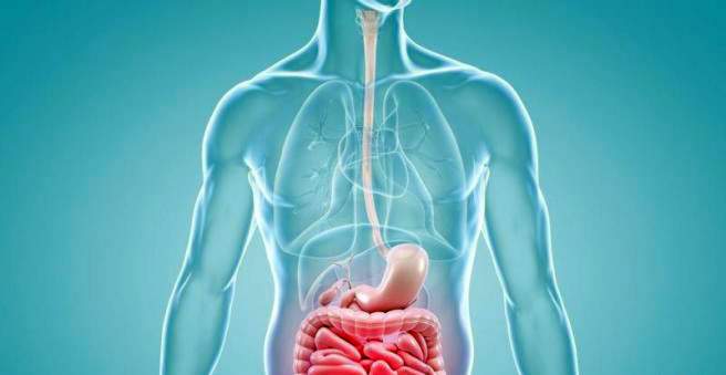
Esophageal varices are varicose veins of the esophagus. The dilated veins occur mainly in advanced cirrhosis. Also in other diseases come as a cause in question, for example, a right heart failure. If esophageal varices break and bleed, there is danger to their lives! Read everything important about symptoms, treatment and prevention of esophageal varices.

How esophageal varices develop
Varicose veins in the esophagus (esophageal varices) are often complications of advanced shrinking (Liver cirrhosis). In this chronic liver disease, functioning liver tissue is increasingly being transformed into functionless connective tissue – it scarred. The more pronounced the scarring, the more impaired the blood circulation in the liver. This can result in esophageal varices and other complications. To understand this, one has to look more closely at the blood flow through the liver:
The blood supply to the liver occurs, inter alia, via the portal vein, This large vessel transports the blood from the intestine (with the absorbed nutrients) and from other abdominal organs (stomach, spleen, etc.) to the liver. It acts as the central metabolic organ in which innumerable substances are constantly being built up, rebuilt and degraded and harmful substances are detoxified. After flowing through the liver, the blood flows through the hepatic veins into the inferior vena cava and on to the right heart.
In liver cirrhosis, however, the increasing scarring of the tissue causes the blood to stop flowing properly through the liver. It builds up in front of the liver in the portal vein. As a result, the pressure inside the vessel increases morbidly: it develops portal hypertension (portal hypertension).
The jammed blood seeks another way to the inferior vena cava, that is, it is formed Collateral circulation, One of them runs from the portal vein via the gastric veins into the lower esophageal veins, and in this way reaches the inferior vena cava. However, because the veins of the esophagus are very thin-walled, they can not withstand the increased blood flow: they expand and “lament” – it produces varicose veins of the esophagus (esophageal varices).
Attention: There are also other diseases that can cause portal vein hypertension and, subsequently, esophageal varices. These include a right heart failure (right heart failure) and a blockage of the portal vein by a blood clot (portal vein thrombosis).
In addition to those esophageal varices, which have caused by other diseases, there are also primary esophageal varices: These are not based on another disease, but on a congenital malformation of the vessels. They are very rare.
Bleeding from esophageal varices
The increased blood flow can dilute the wall of the esophageal vein so much over time that it bursts. Doctors then speak of one esophageal varices, Such bleeding is life-threatening and can lead to death in no time!
Oesophageal varice bleeding occurs in 30 percent of all cirrhotic patients and is one of the leading causes of death in this disease. The further cirrhosis has progressed, the more likely patients are to die from esophageal variceal hemorrhage.
The highest risk of oesophageal variceal hemorrhage is in patients who:
- already had an esophageal variceal hemorrhage
- continue to drink alcohol (the main cause of liver cirrhosis)
- have very large esophageal varices
Esophageal varices: symptoms
Esophageal varices usually do not cause discomfort as long as they are intact. The affected people do not even notice them.
Only when oesophageal varices tear, they make themselves suddenly noticeable: The patients vomit suddenly and gushing a large amount of blood. Due to the blood and fluid loss, symptoms of hypovolemic shock quickly develop. These include, for example, cool and pale skin, drop in blood pressure, rapid heartbeat, flat breathing and impaired consciousness.
Attention: An esophageal variceal hemorrhage must be treated as quickly as possible – there is a high risk of death!
Esophageal varices: diagnosis
Esophageal varices can be detected in an endoscopy, more specifically in a speculum of the esophagus (ostrophagoscopy) or a gastroscopy (gastroscopy). A thin tube is inserted through the mouth into the esophagus (in a gastroscopy even further into the stomach). At its front end sit a light source and small camera. This constantly takes pictures of the inside of the esophagus and plays them on a monitor. Esophageal varices can usually be recognized quite quickly on the images.
If a patient vomits blood, the suspicion of esophageal variceal hemorrhage is near. It can also be another source of bleeding in the upper digestive tract. These include, for example, gastric ulcers (ulcers) and gastritis with damage to the mucous membrane (erosive gastritis).
Esophageal varices: therapy
If esophageal varices are detected at endoscopy, the doctor may consider them precautionary become deserted, Another method to reduce the risk of esophageal variceal hemorrhage is the so-called Rubber band ligation (Varicose ligature): The extended vein is ligated with a small rubber band or with several rubber bands. As a result, it heals and can no longer bleed.
Both measures are possible in the scope of endoscopy: The thin tube allows the doctor to insert the necessary fine instruments into the esophagus.
Esophageal varices bleeding: therapy
If esophageal variceal hemorrhage occurs, action must be taken quickly: the most important emergency measure is to stabilize the patient’s circulation. With a torn esophageal vein, much blood and fluid are lost in a very short time. Therefore, the patient Liquid directly into a vein and if necessary, too blood transfusions administered.
In parallel, attempts are made to stop the bleeding. There are several methods available for this:
First and foremost, the endoscopic one is used for this Rubber band ligation (Varicose ligation, as described above). Additionally or alternatively, the doctor may Medication for hemostasis such as somatostatin or terlipressin. They lower the blood pressure in the portal vein system.
Sometimes, with an esophageal varix hemorrhage, the affected one is also Vessel desolate (as part of an endoscopy).
In massive bleeding can also be a so-called balloon tamponade help: A small, empty balloon is inserted into the lower esophagus and then inflated. This will squeeze the blood vessels, stopping the bleeding. But the method carries some risk. For example, if the balloon is inflated too much, the esophagus may rupture. The balloon can also slip in the direction of the head and block the airways. Because of these risks, balloon tamponade is usually only performed in severe, uncontrollable esophageal varix bleeding.
In the further course, patients often receive precautionary measures antibioticsto prevent a possible bacterial infection.
Since esophageal variceal hemorrhage usually occurs in cirrhosis, one also has to Prevent the risk of liver congestion, Normally, the blood that enters the gastrointestinal tract after bleeding is broken down by the liver cells. Due to cirrhosis of the liver, the liver is unable to do so. Therefore, toxic metabolic products can accumulate. If they get into the head via the blood, they can damage the brain (hepatic encephalopathy). Therefore, the blood that is still present in the esophagus, must be aspirated. In addition, the patient receives lactulose – a light laxative to cleanse the colon.
Prevention of recurrent bleeding
Within ten days of the first esophageal variceal hemorrhage, approximately 35 percent of patients ruptured a varicose vein in the esophagus again. Within a year of the first bleeding, this is even true for 70 percent of patients.
The so-called secondary prophylaxis is therefore very important. It includes measures to prevent recurrence of esophageal varices. Thus, many patients receive a blood pressure lowering drug (such as propranolol) for portal blood pressure. Sometimes, as a precaution, a varicose ligation is performed.
In certain cases, it may also make sense to create a so-called “shunt” (TIPS). In other words, a connection is made between the portal vein and the hepatic veins, which bypasses the scarred tissue of the liver. This prevents the blood from making a detour via the esophageal veins and new ones esophageal varices caused or enlarged existing.