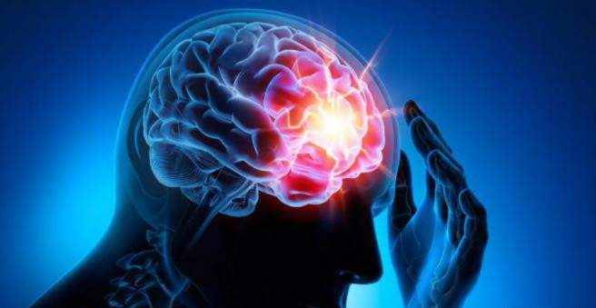
The stroke (apoplexy, stroke) is a sudden circulatory disorder in the brain. She must be treated as soon as possible! Otherwise, so many brain cells die that the patient sustains, or even dies, permanent damage such as paralysis or speech disorder. Read all important information on the topic: What exactly is a stroke and how does it develop? What are the warning signs and what consequences can he have? How is he treated?

Stroke: Short overview
- What is a stroke? A sudden circulatory disorder in the brain
- Important symptoms: Acute muscle weakness, paralysis and numbness in one half of the body, sudden vision and speech disorders, acute and very severe headache, acute dizziness, speech disorders, etc.
- Causes: Low blood flow in the brain, usually due to a blood clot (ischemic stroke), more rarely due to cerebral hemorrhage (haemorrhagic stroke)
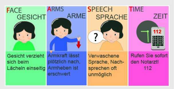
- Stroke test (FAST test): Ask the patient to smile one at a time (F for face), raise both arms simultaneously (A like arms) and repeat a simple sentence (S like speech). If he has any problems, it is probably a stroke and you should quickly alert the ambulance (T as time).
- First aid: Call the emergency doctor (Tel. 112), calm the patient, loosen tight clothing, store upper body (if patient conscious), stable lateral position (in case of unconsciousness), resuscitation measures (if no pulse / no respiration detected)
- Treatment: Stabilization and monitoring of the vital signs, further measures depending on the cause of the stroke (removal of the blood clot by medication or catheter, operation in case of extensive cerebral hemorrhage, etc.), treatment of complications (epileptic seizures, increased intracranial pressure, etc.)
Stroke: description
Stroke is a sudden circulatory disorder in the brain. It is also called apoplexy or apoplexy, stroke, cerebral insult, apoplectic insult or cerebral insult.
The acute circulatory disorder of the brain has the consequence that the brain cells receive too little oxygen and nutrients. As a result, they die. Failures of brain functions can be the result, such as numbness, paralysis, speech or vision disorders. With rapid treatment they can sometimes recede; in other cases they persist permanently. A severe stroke can also be fatal.
Stroke: frequency
Almost 270,000 people in Germany suffer a stroke each year. For about 200,000 of them, it is the first apoplex. The stroke occurs mainly in the elderly. As their proportion in the population is steadily increasing, the number of stroke patients is likely to go up, experts believe.
Stroke in children
Although a stroke usually affects the elderly, it can also occur at a young age. Even unborn children in the womb can already suffer a stroke. Possible causes include, for example, coagulation disorders, cardiovascular diseases. Sometimes an infectious disease triggers a stroke in children.
In Germany, about 300 children and adolescents are diagnosed with apoplexy each year. However, experts suggest that the actual number is much higher because the diagnosis of stroke is more difficult for children. The reason is that the brain maturation is not yet completed and therefore a stroke in children is often only months or years later noticeable. For example, hemiplegia in newborns only occurs after about six months.
Stroke: symptoms
The stroke symptoms depend on which brain region is affected and how severe the stroke is. Very often they show up acute weakness, numbness and paralysis feelings in a body side. This can be recognized, for example, by the fact that the corner of the mouth and the eyelids of one side hang down and / or the patient can no longer move one arm. The left side of the body is affected when the stroke occurs in the right hemisphere, and vice versa. If the patient is completely paralyzed, this indicates a stroke in the brainstem.
Also sudden blurred vision are common stroke symptoms: those affected report, for example, that they can only see blurry or double vision. A sudden, transient visual loss in one eye may also indicate a stroke. Due to the acute visual disturbances, it can happen that the affected person falls or – while driving a car – causes an accident.
A Acute speech disorder can also be a sign of a stroke: Some patients suddenly speak blurred or loud, twisting letters or can not talk. Often, stroke patients can not even understand what they say to them. This is called Language comprehension disorder designated.
Other possible signs of stroke may be sudden dizziness and very severe headache.
You can read more about the signs and symptoms of a stroke in the article Stroke: Symptoms.
Transient ischemic attack (TIA) – the “mini-stroke”
The term “Transient ischemic attack” (short: TIA) refers to a temporary circulatory disorder in the brain. It is an early warning sign for a stroke and is sometimes called a “mini-stroke”.
The TIA is usually caused by tiny blood clots, which temporarily affect the blood flow to a cerebral vessel. The affected person notices this, for example, to temporary speech or vision problems. Sometimes, weakness, paralysis, or numbness in one half of the body may occur for a short time. A temporary confusion or disturbance of consciousness may also occur.
Such TIA symptoms are always sudden and disappear after minutes or a few hours. Nevertheless, one should consult a doctor immediately: If the right therapy is initiated quickly, a “true” stroke can often be prevented.
Everything important about the “mini-stroke” read in the article Transitory ischemic attack.
Stroke: causes and risk factors
Physicians distinguish different stroke causes: The two most common are a reduced blood flow (ischemic stroke) and a cerebral hemorrhage (haemorrhagic stroke). In rare cases, other stroke causes can be identified.
Stroke Cause # 1: Low Perfusion
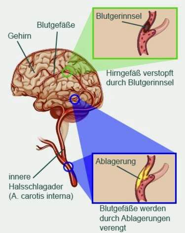
An acute shortage or deficiency (ischaemia) in certain brain regions is the most common of all stroke causes. She is responsible for 80 to 85 percent of all cases of stroke. Doctors speak of one here ischemic stroke or cerebral infarction.
There may be different reasons for the lack of blood flow in certain brain regions. The most important are:
- Blood clots: A blood plug can occlude a cerebral vessel and thus prevent the blood and oxygen supply to a brain region. The clot has often formed in the heart (such as in atrial fibrillation) or in a “calcified” carotid artery and was flushed with the bloodstream into the brain.
- “Vascular calcification” (arteriosclerosis): Cerebral vessels or cerebral vessels in the neck (such as the carotid artery) can “calcify”: deposits on the inner wall constrict a vessel more or even close it completely. The brain area to be supplied then contains too little blood and oxygen.

Particularly serious consequences may be an ischemic stroke in the brainstem (Brainstem infarction). There are vital brain centers, which are responsible for controlling the breathing, the circulation and the consciousness. An example of a brain stem infarction is the basilar artery thrombosis, ie the occlusion of the basilar artery in the brainstem: in severe cases, it causes complete paralysis of all extremities (tetraparesis) and coma or leads to immediate death.
Stroke Cause # 2: Cerebral Hemorrhage
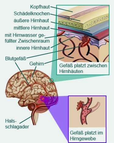
In about 15 to 20 percent of all strokes, head bleeding is the cause. Stroke by such cerebral hemorrhage will also hemorrhagic stroke called. The bleeding can occur in different places:
- Bleeding in the brain: This suddenly bursts a vessel directly in the brain, and blood enters the surrounding brain tissue. The trigger of this so-called intracerebral hemorrhage is usually hypertension. Other illnesses, drug abuse, and the rupture of congenital vascular malformation (such as aneurysm) in the brain can cause brain bleeding. Sometimes the cause remains unclear.
- Bleeding between the meninges: The stroke arises here through a bleeding in the so-called subarachnoid space: This is the cerebral water-filled gap between middle meninges (arachnoid) and inner meninges (pia mater). The cause of such a subarachnoid hemorrhage is usually a spontaneously burst aneurysm (congenital vascular malformation with an outflow of the vessel wall).

Rare stroke causes
Stroke can have other causes, especially in younger people than a reduced blood flow or cerebral hemorrhage. For example, in some patients the stroke is based on one Inflammation of vessel walls (Vasculitis). Such vasculitis occurs in the context of autoimmune diseases such as giant cell arteritis, Takayasu’s arteritis, Behcet’s disease and systemic lupus erythematosus.
Other rare stroke causes are for example Fat and air embolisms: In this case, fat droplets or invading air clog a cerebral vessel, resulting in cerebral infarction. Fat embolism can occur with heavy bone fractures when high-fat bone marrow is flushed into the blood. Air embolism can be a very rare complication of open heart surgery, thorax or neck.
Congenital coagulation disorders and the Formation of blood clots in the veins are also among the rare stroke causes.
Stroke: risk factors
Stroke does not arise out of nothing. Various factors can contribute to its creation. Some of these stroke risk factors can not be influenced. This includes that Age: The risk of a stroke increases with the years of life. Also not influenceable is a genetic predispositionfor a stroke.
There are also many risk factors that can be specifically reduced. This includes, for example High blood pressure (hypertension): It leads to “vascular calcification” (arteriosclerosis), which means that deposits form on the inner wall of the vessels. As a result, the vessels are increasingly close. This favors a stroke. The more severe the hypertension, the more likely a stroke will be.
An avoidable risk factor for a stroke is too Smoke: The more cigarettes a person smokes a day and the more years the smoking career has lasted, the higher the risk of stroke. There are several reasons for this:
Among other things, smoking promotes vascular calcification (arteriosclerosis) and lipid metabolism disorders – both are further risk factors for stroke. In addition, smoking causes the vessels to contract. The resulting increase in blood pressure favors a stroke.
Smoking also reduces the amount of oxygen that can be transported by the red blood cells (erythrocytes). The tissues and organs get thereby less oxygen, so also the brain. This then signals the bone marrow to produce more red blood cells for oxygen transport. Due to the increase in erythrocytes but the blood is “thickened”. As a result, it flows worse through the still narrowed vessels.
Last but not least, smoking increases the blood’s ability to coagulate – especially because the platelets become stickier. This helps to form clots that can clog a vessel. If this happens in the brain, it results in an ischemic stroke.
It is worthwhile to stop smoking. Just five years after the cessation of smoking, someone has the same stroke risk as people who have never smoked.
Other important risk factors for stroke include:
- Alcohol: High alcohol intake – whether regular or rare – increases the risk of stroke. Above all the danger for a brain haemorrhage increases. In addition, regular consumption of alcohol involves further health risks (addictive potential, increased risk of cancer).
- Overweight: Obesity increases the risk of many different diseases. In addition to diabetes and hypertension, this includes stroke.
- Lack of exercise: Possible consequences are obesity and hypertension. Both favors a stroke.
- Dyslipidemia: LDL (“bad”) cholesterol and other blood lipids are part of the deposits that settle on the inner walls of vessels in arteriosclerosis. High blood lipid levels (such as high cholesterol levels) increase the risk of stroke via arteriosclerosis.
- Diabetes mellitus (diabetes): Diabetes damages the blood vessel walls, causing them to thicken. This affects the blood flow. which affects the blood flow. In addition, diabetes aggravates an existing arteriosclerosis. Overall, diabetics have a two to three times higher risk of stroke than people who are not diabetic.
- Atrial Fibrillation: This cardiac arrhythmia increases the risk of stroke because it easily forms blood clots in the heart. These can – entrained by the blood stream – in the brain clog a vessel (ischemic stroke). This risk is even greater if, in addition, other heart diseases exist, such as coronary heart disease (CHD) or heart failure.
- Other cardiovascular diseases: Other cardiovascular diseases such as smoker’s leg (PAD) and “impotence” (erectile dysfunction) increase the risk of stroke.
- Narrowed carotid artery (carotid stenosis): It is usually based on vascular calcification (arteriosclerosis) and often causes no complaints for a long time. Possible early symptom is a TIA (Transient Ischemic Attack). Whether asymptomatic or not – the carotid stenosis increases the risk of ischemic stroke (cerebral infarction).
- Aura migraines: Low blood flow stroke often occurs in people who suffer from a migraine with aura. The headache is preceded by neurological symptoms such as visual or sensory disorders. The exact relationship between aura migraine and stroke is not yet known. Especially women are affected.
- Hormone preparations for women: Taking the contraceptive pill increases the risk of stroke. This is especially true in women with other risk factors such as high blood pressure, smoking, obesity or aura migraine. The use of hormone supplements during menopause (hormone replacement therapy, HET) increases the risk of stroke.
Pediatric stroke: causes
Stroke in children is rare but occurs. While adult lifestyle factors and lifestyle diseases (smoking, atherosclerosis, etc.) are the main cause of stroke, children have other stroke causes. These include, for example, an inherited tendency to clot, disorders of the red blood cells (such as sickle cell anemia) and connective tissue diseases (such as Fabry disease). Also autoimmune diseases of the blood vessels as well as heart diseases are possible stroke causes in children.
Stroke: examinations and diagnosis
Whether severe or mild stroke – every stroke is an emergency! Already on mere suspicion you should Call the emergency doctor immediately (Tel. 112)! With the FAST Test You can easily and quickly check for suspected stroke. The stroke test works as follows:
- F as “face”: Ask the patient to smile. If the face is distorted on one side, this indicates a hemiplegia due to a stroke.
- A like “arms”: Ask the patient to extend their arms forward at the same time, turning their palms upwards. If he has problems, there is probably an incomplete paralysis of one half of the body as a result of a stroke.
- S as “speech” (language): Ask the patient to repeat a simple sentence. If he is unable to do so or if his voice is blurred, there is probably a speech disorder as a result of a stroke.
- T as “time”: Call the ambulance immediately!

The emergency physician will check, among other things, the patient’s awareness, blood pressure and heart rate. If he is conscious, the doctor can ask him about what is happening and symptoms that occur (such as blurred vision, numbness or paralysis).
After admission to the hospital, a neurologist is the specialist responsible for suspected stroke. He leads one neurological examination by. He examines, for example, coordination, language, vision, touch and reflexes of the patient.
As a rule, one will be immediately Computed tomography of the head (cranial computed tomography, cCT). The examination is often supplemented by a vessel imaging (CT angiography) or a blood perfusion measurement (CT perfusion). The pictures inside the skull show whether a vascular occlusion or a cerebral hemorrhage is responsible for the stroke of the brain. You can also see its location and extent.
Sometimes, instead of computed tomography, one becomes Magnetic Resonance Imaging (MRI) stated. It can also be combined with a vascular presentation or blood flow measurement.
In some patients, a separate X-ray examination of the vessels (angiography) carried out. Vascular imaging is important to detect, for example, vascular malformations (such as aneurysms) or vascular leaks.
To clarify a stroke may also be a special Ultrasound (Doppler and duplex sonography) of the brain-supplying vessels such as the carotid artery. At the same time, the doctor can detect whether there is calcification (arteriosclerotic deposits) on the inner wall of the vessel. They can be the site of a blood clot that has been carried away by the bloodstream and caused the stroke.
A Ultrasound examination of the heart cavities (Echosonography) can show heart disease, which promotes the formation of blood clots, for example, deposits on the heart valves. Sometimes blood clots are found in the heart caves. They could cause another stroke. Therefore, the patient must be given blood-thinning medication, which dissolve the blood clots.
Another important cardiac examination after a stroke is the Electrocardiography (ECG), This is the measurement of the electrical heart currents. Sometimes it is also used as a long-term measurement (24-hour ECG or long-term ECG). Based on the ECG, the doctor can detect any cardiac arrhythmia. They are also an important risk factor for an ischemic insult.
Another important factor in stroke diagnosis is one blood test, For example, blood count, blood clotting, blood sugar, electrolytes and kidney values are determined.
These studies are not only intended to confirm the suspicion of apoplexy and to clarify it further. They also help to detect possible complications early on. These include, for example, blood pressure crises, an additional myocardial infarction, pneumonia caused by inhalation of food residues (aspiration pneumonia) and kidney failure.
Stroke: treatment
In the case of stroke treatment, every minute counts because the principle applies “Time is brain”: Brain cells that are not adequately blooded or squeezed by increased intracranial pressure, depending on the type of stroke, die rapidly. Stroke patients should therefore receive medical attention as soon as possible!
Stroke: first aid
For any suspected stroke you should immediately alert the ambulance (emergency number 112)! Until this happens, you should calm the patient down. Store your upper body slightly raised and open tight clothing (such as a collar or tie). That makes breathing easier. Do not give him anything to eat or drink!
If the patient is unconscious but is breathing, you should put him in stable lateral position (on the paralyzed side). Regularly check his breathing and his pulse.
If you can not find any signs of breathing, you should immediately turn the affected person on his back and start the cardiopulmonary resuscitation (chest compressions and possibly mouth-to-mouth resuscitation).
For acute acute treatment in each stroke, it is important to monitor the vital signs and other important parameters and to stabilize them if necessary. These include, for example, breathing, blood pressure, heart rate, blood sugar, body temperature, brain and kidney function as well as water and electrolyte balance. Further measures depend on the type of stroke and possible complications.
Stroke: treatment for cerebral infarction
Most cerebral infarctions (ischemic strokes) are caused by a blood clot that clogs a cerebral vasculature. This must be quickly eliminated to restore the circulation in the affected area of the brain and rescue nerve cells from destruction. The blood clot can either dissolve with a drug (lysis therapy) or mechanically eliminate (thrombectomy). Both methods can also be combined with each other.
Lysis therapy
In the so-called Systemic lysis The stroke patient receives an infusion into a vein a drug that can dissolve blood clots (thrombolytic). The active substance rtPA (“recombinant tissue plasminogen activator”) is used. This activates an enzyme in the body that breaks down blood clots. This form of lysis therapy is approved up to 4.5 hours after cerebral infarction. The sooner lysis begins within this time frame, the higher the chances of success.
If more than about 4.5 hours have passed, the clot can hardly dissolve any more medicament. Nevertheless, in certain cases systemic lysis with rtPA can also be carried out up to 6 hours after the occurrence of the stroke symptoms – as an individual therapeutic attempt.
The lysis therapy must not be carried out in the event of a stroke due to cerebral hemorrhage. That could aggravate the bleeding. In certain other situations lysis therapy is not recommended, for example in uncontrollable hypertension.
In addition to the systemic lysis therapy, there are also the local lysis (intra-arterial thrombolysis). This involves advancing a catheter over an artery to the site of vascular occlusion in the brain and directly injecting a clot-dissolving drug (such as pro-urokinase). However, the local lysis therapy is only suitable in very specific cases (such as brain stem infarction).
thrombectomy
Another form of stroke treatment is based on the mechanical removal of the blood clot: In the so-called thrombectomy, a thin catheter is advanced under X-ray control over an artery in the groin to the clot in the brain. This is then removed with suitable fine instruments. Thrombectomy should be performed as soon as possible after the onset of stroke symptoms.
Combination of thrombolysis and thrombectomy
There is also the possibility to combine both procedures – the dissolution of the blood clot in the brain with a drug (thrombolysis) and the mechanical removal of the clot by catheter (thrombectomy).
Stroke: treatment for cerebral hemorrhage
If the stroke is triggered by a smaller cerebral hemorrhage, usually enough conservative stroke treatment out. Patients need to keep bed rest and avoid any activities that increase the pressure in the head. This includes strong pressing during bowel movements. Therefore, the patients receive laxatives.
It is also very important to monitor the blood pressure and to treat it as needed: too high a pressure intensifies the bleeding, too low a pressure could lead to a deficient circulation of brain tissue.
Cerebral hemorrhage, which is more extensive and does not stop by itself, is usually one surgery necessary. However, the decision to undergo surgery depends on various factors, such as the location and size of the bleeding, the age and general condition of the patient, as well as any comorbidities. During surgery, the skull is opened to eliminate the bruise (Hämatoevakuation) and the source of bleeding as possible to close.
Stroke: treatment of complications
As needed, stroke treatment includes further measures, especially if complications occur.
Increased intracranial pressure
In a very large cerebral infarction, the brain can swell (brain edema). Because the space in the bony skull is limited, the intracranial pressure increases as a result. Nerve tissue can be squeezed and damaged irreversibly.
Even with a larger cerebral hemorrhage, the pressure in the skull can rise due to the escaping blood. When blood enters the brain ventricles filled with nerve water, the nerve water can also build up – a hydrocephalus develops. This also increases the intracranial pressure dangerous.
Whatever the reason for increased intracranial pressure, it must be lowered. For this purpose, for example, the head and upper body of the patient are stored high. Also useful is the administration of dehydrating infusions or the discharge of nerve water via a shunt (for example into the abdominal cavity). For relief, a part of the cranial bone can be temporarily removed and later reinserted (relief craniotomy). Eliminating bruising on cerebral hemorrhages also reduces the pressure in the skull.
Vascular spasms (vasospasm)
In a stroke caused by bleeding between the meninges (subarachnoid hemorrhage), there is a risk that the vessels constrict convulsively. Through these vascular spasms (vasospasms), the brain tissue can no longer be sufficiently supplied with blood. Then an additional ischemic stroke can occur. Vascular spasms must therefore be treated with medication.
Epileptic seizures and epilepsy
Stroke is very often the reason for new epilepsy in elderly patients. An epileptic seizure can occur within the first hours after the stroke, but also days or weeks later. Epileptic seizures can be treated medically (with antiepileptic drugs).
lung infection
The most common complications after a stroke include bacterial pneumonia. Particularly high is the risk in patients suffering from dysphagia as a result of stroke: If swallowed, food particles may enter the lungs and cause pneumonia (aspiration pneumonia). Antibiotics are given for prevention and treatment. Stroke patients with dysphagia may also be fed artificially (via a probe). This lowers the risk of pneumonia.
Urinary tract infections
In the acute phase after a stroke patients often can not leave water (urinary retention or urinary retention). Then a bladder catheter must be placed repeatedly or permanently. Both urinary tract and permanent catheters promote a urinary tract infection after a stroke. Treatment is with antibiotics.
Rehabilitation after stroke
The medical rehabilitation after stroke aims to help a patient to return to his old social and possibly also professional environment. For this purpose, for example, with suitable training methods is attempted to reduce functional restrictions such as paralysis, speech and speech disorders or visual disturbances.
In addition, the rehabilitation after stroke should put a patient back in the position to cope with his everyday life as much as possible independently. This includes, for example, washing yourself, dressing or preparing a meal. Sometimes there are physical limitations (such as a paralyzed hand) that make it difficult or impossible to do some manipulation or movement. Then affected people learn in stroke rehabilitation solution strategies and dealing with suitable tools (such as bathtub lift).
Inpatient or outpatient
A neurological rehabilitation can be especially in the beginning after a stroke stationary take place, for example in a rehab clinic. The patient receives an individual treatment concept and is supervised by an interdisciplinary team (doctors, nurses, occupational therapists and physiotherapists etc.).
In the semi-stationary rehabilitation The stroke patient comes to the rehabilitation ward during the day for his therapy hours during the day. He lives at home.
If interdisciplinary care is no longer needed, but the patient still has physical limitations in certain areas, one helps outpatient rehabilitation further. The respective therapist (such as occupational therapist, speech therapist) so comes regularly to the stroke patient home to practice with him.
Motor rehabilitation
The most common complications after a stroke include sensorimotor dysfunction. Darunter versteht man ein gestörtes Zusammenspiel von sensorischen Leistungen (Sinneseindrücken) und motorischen Leistungen (Bewegungen). Meist handelt es sich dabei um die unvollständige Lähmung in einer Körperhälfte (Hemiparese). Verschiedene Therapieformen können helfen, solche sensomotorischen Störungen zu verbessern:
So wird bei einer Halbseitenlähmung besonders oft das Bobath-Konzept angewendet: Die gelähmte Körperpartie wird beharrlich gefördert und stimuliert. Beispielsweise wird der Patient nicht gefüttert, sondern gemeinsam mit ihm und dem beeinträchtigten Arm wird der Löffel zum Mund geführt. Auch bei jeder anderen Aktivität im Alltag muss das Bobath-Konzept umgesetzt werden – mithilfe von Ärzten, Pflegekräften, Angehörigen und allen anderen Betreuern. Mit der Zeit kann sich das Gehirn so umorganisieren, dass gesunde Hirnteile nach und nach die Aufgaben der geschädigten Hirnareale übernehmen.
Ein anderer Ansatz ist dieVojta-Therapie.Sie beruht auf der Beobachtung, dass viele Bewegungen des Menschen reflexartig ablaufen, so etwa das reflexartige Greifen, Krabbeln und Umdrehen im Babyalter. Diese sogenannte Reflexlokomotion ist auch beim Erwachsenen noch präsent, wird aber normalerweise von der bewussten Bewegungskontrolle unterdrückt.
Bei der Vojta-Methode werden solche Reflexe gezielt ausgelöst. Der Therapeut reizt zum Beispiel bestimmte Druckpunkte am Rumpf des Patienten, was spontane Muskelreaktionen hervorruft (zum Beispiel richtet sich der Rumpf automatisch gegen die Schwerkraft auf). Bei regelmäßigem Training sollen auf diese Weise gestörte Nervenbahnen sowie bestimmte Bewegungsabläufe reaktiviert werden.
Kognitiv therapeutische Übungen nach Perfetti eignen sich besonders bei neurologischen Störungen und Halbseitenlähmung. Der Patient soll die Bewegungsabläufe neu erlernen und die verlorene Bewegungskontrolle zurückgewinnen. Dazu muss er zunächst Bewegungen erspüren: Mit geschlossenen Augen oder hinter Sichtschutz werden gezielte Bewegungen etwa mit der Hand oder dem Fuß ausgeführt, die der Patient bewusst spüren soll. Anfangs führt der Therapeut noch die Hand oder den Fuß des Patienten, um falsche Muster zu vermeiden. Später führt der Patient die Bewegungen selbst aus, wird aber vom Therapeuten noch unterstützt oder korrigiert. Schließlich lernt der Schlaganfall-Patient, schwierigere Bewegungsabläufe allein auszuführen und Störungen über das Gehirn zu kontrollieren.
The „Forced-use“ Therapie (engl. für „erzwungener Gebrauch“) wird auch „Constrained Induced Movement“ genannt. Sie wird in der Regel eingesetzt, um einen teilgelähmten Arm und die dazugehörige Hand zu trainieren, manchmal auch die unteren Gliedmaßen. Bei manchen der Betroffenen regeneriert sich das geschädigte Hirnareal mit der Zeit soweit, dass die erkrankte Körperpartie nach und nach wieder funktionstüchtiger wird. Das Problem: Der Betroffene hat inzwischen komplett verlernt, die kranken Gliedmaßen zu bewegen, und setzt sie daher kaum oder gar nicht ein.
Hier setzt die „Forced-use“ Therapie an: Indem sich der Patient zum Einsatz der betroffenen Gliedmaße zwingt, soll sie weitgehend reaktiviert werden. Dafür notwendig ist ein anstrengendes Training der teilgelähmten Gliedmaße. Beispielsweise üben die Teilnehmer in stetiger Wiederholung spezielle Bewegungen ein. Durch den häufigen Gebrauch erweitert sich das Hirnareal, das für den betreffenden Körperteil zuständig ist, und es entstehen neue Nervenverbindungen.
Rehabilitation bei Schluckstörungen
Schluckstörungen (Dysphagien) sind weitere häufige Folgen eines Schlaganfalls. Mit der richtigen Therapie soll der Betroffene die Fähigkeit zu essen und zu trinken wiedererlangen. Gleichzeitig soll das Risiko, sich zu verschlucken, gesenkt werden. Um dies zu erreichen, gibt es drei verschiedene Therapieverfahren, die auch miteinander kombinierbar sind:
- Restituierende (wiederherstellende) Verfahren: Mithilfe von Stimulations-, Bewegungs- und Schluckübungen wird versucht, die Schluckstörung zu beseitigen. Das kann etwa gelingen, indem andere Hirnareale die Aufgabe des geschädigten Hirnbereichs ganz oder teilweise übernehmen.
- Kompensatorische Verfahren: Veränderungen der Haltung und Schluckschutz-Techniken sollen das Risiko senken, dass sich der Patient verschluckt. Wenn nämlich Nahrungsreste oder Flüssigkeiten in der Lunge landen, kommt es zu Hustenattacken, Erstickungsanfällen oder Lungenentzündung (Aspirationspneumonie).
- Adaptierende Verfahren: Die Kostform wird so angepasst, dass Patienten mit Schluckstörungen das Essen und Trinken leichter fällt. ZUm Beispiel werden Speisen püriert und Getränke angedickt. Als Unterstützung kommen Therapiehilfen wie spezielle Trinkbecher oder spezielles Besteck zum Einsatz.
Kognitive Rehabilitation
Die kognitive Reha nach Schlaganfall versucht, gestörte kognitive Funktionen wie Sprache, Aufmerksamkeit oder Gedächtnis zu verbessern. Wie bei der Therapie von Schluckstörungen kann auch hier die Rehabilitation auf Restitution (Wiederherstellung), Kompensation oder Adaptation (Anpassung) abzielen. Zum Einsatz kommen ganz unterschiedliche Therapieverfahren.
So können etwa bei Aufmerksamkeits-, Gedächtnis- und Sehstörungen zum Beispiel computergestützte Trainingsverfahren hilfreich sein. Bei Gedächtnisstörungen können Lernstrategien die Gedächtnisleistung verbessern und Hilfsmittel wie ein Tagebuch eine Kompensationsmöglichkeit bieten. In bestimmten Fällen kommen auch Medikamente zum Einsatz.
Vorbeugung eines weiteren Schlaganfalls
Bei jedem Patienten müssen nach Möglichkeit bestehende Ursachen und Risikofaktoren für den Schlaganfall beseitigt oder zumindest reduziert werden. Das hilft, einem weiteren Hirnschlag vorzubeugen (Sekundärprophylaxe). Zu diesem Zwecke müssen oft lebenslang Medikamente eingenommen werden. Auch nicht-medikamentöse Maßnahmen sind wichtig für die Sekundärprophylaxe.
„Blutverdünner“ (Thrombozytenaggregationshemmer): Nach einem Schlaganfall durch Minderdurchblutung oder einer TIA (“Mini-Schlaganfall”) erhalten die meisten Patienten sogenannteThrombozytenfunktionshemmer. Dazu zählen zum Beispiel Acetylsalicylsäure (ASS) und Clopidogrel. Diese „Blutverdünner“ verhindern, dass Blutplättchen zu einem Pfropf verklumpen, der dann vielleicht erneut ein Gefäß verstopft. Die Medikamente sollten nach Möglichkeit lebenslang eingenommen werden.
Übrigens: ASS kann als Nebenwirkung ein Magen-oder Zwölffingerdarmgeschwür verursachen. Betroffene Patienten müssen deshalb oft zusätzlich zu ASS einen sogenannten Protonenpumpenhemmer („Magenschutz“) einnehmen.
Gerinnungshemmer (Antikoagulanzien): Ein Schlaganfall durch Minderdurchblutung (ischämischer Schlaganfall) oder eine TIA (“Mini-Schlaganfall”) entsteht oft als Folge von Vorhofflimmern. Bei dieser Herzrhythmusstörung bilden sich sehr leicht Blutgerinnsel im Herz, die dann vom Blutstrom mitgerissen werden und ein Gefäß im Gehirn verstopfen können. Damit das nicht nochmal passiert, erhalten Schlaganfall-Patienten mit Vorhofflimmern gerinnungshemmende Medikamente in Tablettenform (orale Antikoagulanzien). Diese Medikamente blockieren den komplizierten Prozess der Blutgerinnung und damit die Gerinnselbildung.
Cholesterinsenker: Eine der Hauptursachen von Schlaganfall ist die Gefäßverkalkung (Arteriosklerose). Bestandteil der Kalkablagerungen an der Gefäßinnenwand ist Cholesterin. Nach einem Schlaganfall durch Minderdurchblutung (ischämischer Apoplex) sowie nach einem “Mini-Schlaganfall” (TIA) erhalten Patienten deshalb meistens cholesterinsenkende Medikamente aus der Gruppe der Statine (CSE-Hemmer). Diese verhindern, dass eine bestehende Arteriosklerose weiter fortschreitet.
Bei einem Schlaganfall durch Hirnblutung werden Cholesterinsenker nur bei Bedarf und nach sorgfältiger Nutzen-Risiko-Abwägung verordnet.
Blutdrucksenker (Antihypertensiva): Bluthochdruckpatienten müssen nach einem ischämischen Schlaganfall oder einer TIA langfristig blutdrucksenkende Medikamente einnehmen. Das soll einen erneuten Hirnschlag verhindern. Der behandelnde Arzt entscheidet im Einzelfall, welcher Blutdrucksenker am besten geeignet ist (ACE-Hemmer, Betablocker etc.) und welcher Blutdruck-Zielwert angestrebt wird.
Nicht-medikamentöse Maßnahmen: Manche Risikofaktoren für einen erneuten Schlaganfall lassen sich (unterstützend) auch mit nicht-medikamentösen Maßnahmen verringern. Empfohlen werden zum Beispiel der Abbau von Übergewicht, regelmäßige Bewegung, eine ausgewogene Ernährung mit wenig tierischen Fetten und der Verzicht auf Nikotin und Alkohol. Ein solcher Lebensstil hilft unter anderem, zu hohe Blutdruck- und Cholesterinwerte in den Griff zu bekommen. Das senkt wesentlich das Risiko für einen weiteren Schlaganfall.
Schlaganfall: Stroke Unit
Unter dem Begriff “Stroke Unit” versteht man eine spezielle Abteilung in einem Krankenhaus, deren Mitarbeiter auf die Diagnose und Akutbehandlung von Menschen mit Hirnschlag spezialisiert sind. Die Betreuung auf einer solchen “Schlaganfall-Station” verbessert nachweislich die Überlebenschancen der Patienten und senkt das Risiko für bleibende Schäden.
Die Patienten bleiben im Schnitt etwa drei bis fünf Tage in der “Stroke Unit”. Danach werden sie je nach Bedarf auf eine andere Station (neurologische Station, Allgemeinstation) verlegt oder direkt in eine Rehabilitationseinrichtung überwiesen.
Es gibt in Deutschland mittlerweile mehr als 280 “Stroke Units”. Sie werden von der Deutschen Schlaganfall-Hilfe zertifiziert.
Mehr zum Thema erfahren Sie im Beitrag Stroke Unit.
Schlaganfall: Krankheitsverlauf und Prognose
Allgemein gilt: Die Hirnschädigung durch einen Schlaganfall ist umso schwerwiegender, je größer das betroffene Blutgefäß ist, das verstopft wurde oder geplatzt ist. Allerdings können sich in besonders empfindlichen Gehirnregionen wie beispielsweise dem Hirnstamm auch schon kleine Schäden verheerend auswirken.
Rund ein Fünftel (20 Prozent) aller Hirnschlag-Patienten verstirbt innerhalb der ersten vier Wochen. Im Laufe des ersten Jahres sterben mehr als 37 Prozent der Betroffenen. Insgesamt ist der Schlaganfall nach Herz- und Krebserkrankungen die dritthäufigste Todesursache in Deutschland.
Von jenen Schlaganfall-Patienten, die nach einem Jahr noch leben, trägt etwa die Hälfte bleibende Schäden davon und ist dauerhaft auf fremde Hilfe angewiesen. In Deutschland sind das fast eine Million Menschen.
Ein Schlaganfall bei Kindern hat sehr gute Heilungschancen. Es gibt gute Behandlungsmöglichkeiten für die kleinen Patienten, sodass die meisten von ihnen nach einiger Zeit wieder ein normales Leben führen können. Nur bei ungefähr zehn Prozent aller betroffenen Kinder hinterlässt der stroke eine größere Beeinträchtigung.
Schlaganfall: Folgen
Viele Patienten haben nach einem Schlaganfall bleibende Beeinträchtigungen. Dazu zählen zum Beispiel Bewegungsstörungen wie ein unsicherer Gang oder eine Halbseitenlähmung. Manche Patienten haben Schwierigkeiten, ihre Bewegungen zu koordinieren (etwa beim Schreiben) oder komplexe Bewegungen auszuführen (wie etwa das Öffnen eines Briefes).
Zu den möglichen Schlaganfall-Folgen gehören auch Sprach- und Sprechstörungen: Bei einer Sprachstörung haben Betroffene Probleme, ihre Gedanken zu formulieren (mündlich oder schriftlich) und/oder zu verstehen, was andere ihnen sagen. Dagegen ist bei einer Sprechstörung das motorische Artikulieren von Wörtern beeinträchtigt.
Weitere häufige Folgen eines Schlaganfall sind zum Beispiel Störungen der Aufmerksamkeit und des Gedächtnisses sowie Seh- und Schluckstörungen. Mehr darüber lesen Sie im Beitrag Schlaganfall: Folgen.
Leben mit Schlaganfall
Nach einem Schlaganfall ist oft nichts mehr so, wie es vorher war. Folgeschäden wie zum Beispiel Seh- und Sprachstörungen sowie Halbseitenlähmung können das ganze Alltagsleben beeinflussen. Beispielsweise kann nach einem Hirnschlag die Fahrtüchtigkeit so stark beeinträchtigt sein, dass sich Patienten besser nicht mehr hinters Lenkrad setzen. Aber auch, wer scheinbar fit ist, sollte freiwillig die Führerscheinstelle über den Schlaganfall informieren und ein ärztliches Gutachten einreichen. Eventuell verlangt die Behörde zusätzliche Fahrstunden oder ein Umrüsten des Fahrzeugs.
Bei jüngeren Menschen stellt sich nach einem Schlaganfall die Frage, ob eine Rückkehr in den Beruf möglich ist oder eine Umschulung notwendig wird. Auch Urlaubsreisen erfordern nach einem Hirnschlag oft besondere Kompromisse und Anpassungen.
Das Leben nach einem Schlaganfall stellt Angehörige ebenfalls vor Herausforderungen. Es geht darum, den Patienten im Alltag möglichst zu unterstützen, ihm aber auch nicht alles abzunehmen.
Mehr über die Herausforderungen des Alltaglebens nach einem Hirnschlag lesen Sie im Beitrag Leben mit Schlaganfall.
Schlaganfall vorbeugen
Verschiedenste Risikofaktoren tragen zur Entstehung eines Schlaganfalls bei. Viele davon lassen sich gezielt reduzieren oder sogar ganz beseitigen. Das beugt einem Hirnschlag wirksam vor.
Wichtig ist zum Beispiel eine balanced nutrition mit viel Obst und Gemüse. Dagegen sollten Sie Fett und Zucker nur in Maßen zu sich nehmen. Mit dieser gesunden Kost beugen sie einer Gefäßverkalkung (Arteriosklerose) vor – diese zählt zu den Hauptursachen von Schlaganfall.
Regelmäßige Bewegung und Sport halten die Gefäße ebenfalls gesund und beugen einem Schlaganfall vor. Wenn Sie übergewichtig sind, sollten Sie abnehmen, Überschüssige Kilos erhöhen nämlich das Risiko für Bluthochdruck und Arteriosklerose. Beides begünstigt einen Schlaganfall.
Ein weiterer guter Tipp, um einem Hirnschlag vorzubeugen, ist der Verzicht auf Nikotin und Alkohol.
Mehr darüber, wie Sie das Schlaganfall-Risiko senken können, lesen Sie im Beitrag Schlaganfall vorbeugen.
Additional information
Book recommendations:
- Schlaganfall: Das Leben danach: Experten-Tipps für Menschen mit Schlaganfall und anderen Schäden des zentralen Nervensystems (Rainer Schulze-Muhr, CreateSpace Independent Publishing Platform, 2017)
- Als mich der Schlag traf: Nach einem Schlaganfall zurück ins Leben (Gabo, W. Zuckschwerdt Verlag, 2013)
guidelines:
- S1-Leitlinie “Akuttherapie des ischämischen Schlaganfalls” der Deutschen Gesellschaft für Neurologie (2012)
- S3-Leitlinie “Sekundärprophylaxe ischämischer Schlaganfall und transitorische ischämische Attacke” der Deutschen Schlaganfall-Gesellschaft und der Deutschen Gesellschaft für Neurologie (2015)
- S2k-Leitlinie “Akuttherapie des ischämischen Schlaganfalls – Ergänzung 2015: Rekanalisierende Therapie” der Deutschen Gesellschaft für Neurologie (2016)
Selbsthilfegruppen
Stiftung Deutsche Schlaganfall-Hilfe
https://www.schlaganfall-hilfe.de//adressen-selbsthilfegruppen