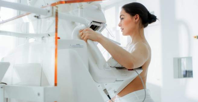
Mammography is an x-ray examination of the breast (lat. Mamma). It serves to clarify changes in breast tissue (such as nodules) that could be breast cancer. In addition, women between the ages of 50 and 69 are invited to routine mammography every two years as part of mammography screening. Read here how the investigation works, from when a mammography makes sense and what risks it holds.

What is mammography?
Mammography is an X-ray examination of the breast for the early detection of breast cancer (mammary carcinoma) or its precursors. These include small calcifications (microcalcifications), nodules, thickening, asymmetries or disturbances in the tissue architecture of the breast.
In the analog mammography The X-ray image is created classically on an exposed film foil. The newer digital mammography allows an electronic storage of the image on a computer, so that certain area can be enlarged and reworked if necessary. Even a three-dimensional image of the breast can be made. This can help the doctor to better evaluate some tissue areas.
Mammography: (Ab) when does it make sense?
(From) when a mammogram makes sense, is still discussed by many experts critical. It is important to weigh the benefits of the study – its high rate of detection of breast cancer – against the risks and disadvantages (see below). In Germany the current situation is as follows:
at Women before the age of 50 Mammography is usually performed only if there is a specific suspicion of breast cancer – for example, because a suspicious node in the chest is palpable. However, if a woman of this age group has an increased breast cancer risk (for example, because her mother or sister is already suffering from breast cancer), the doctor can also, as a precaution, take X-rays of her chest routinely at certain intervals.
at Women between 50 and 69 years then a routine mammographic exam is recommended (Mammography screening). Breast cancer is particularly common in this period of life. Therefore women of this age group are invited every two years as a precautionary measure for mammography. The costs are covered by the health insurance companies (statutory cancer screening program).
at Women over 70 years Recommendations on breast cancer screening (such as mammography) are based on several factors: the individual’s risk of cancer, general health and individual life expectancy are considered.
Women at increased breast cancer risk
Some women are at an increased risk of developing breast cancer, for example because their mother or sister is already suffering from breast cancer or breast cancer risk genes have already been definitively detected in the woman’s genome. Then it may be useful to perform a mammogram even before the age of 50 routinely – often supplemented by a magnetic resonance imaging (magnetic resonance imaging, MRI) of the breast. Such intensified cancer screening depends on the individual risk profile of a woman at increased risk of breast cancer.
How does the mammography work?
The mammography is one outpatient examination, The first time you visit, you will need to complete a questionnaire that collects personal data, pre-existing conditions and, most importantly, breast cancer in the family. The doctor will also talk to you personally about important background information (anamnesis interview).
Tip: You should not apply a deodorant before mammography, as this may affect the informative value of the X-ray image.
For the mammography itself, you need to completely clear your upper body and remove any jewelry that might obscure the breast tissue (necklaces, breast piercings, etc.). Then your breasts are carefully stretched out and compressed as flat as possible between two Plexiglas plates. It may be painful for these mammographic steps. Then the breast tissue is X-rayed. It will be usually two X-ray images from different directions made: from top to bottom (cranio-caudal) and diagonally from the middle to the side (mediolateral oblique).
In good radiological practices, the evaluation of X-ray images is based on the Four-eyes principle, This means that two X-ray specialists (radiologists) examine the images independently of each other. If the findings differ from each other, a new mammogram or another examination such as magnetic resonance imaging (MRI) or a galactography is performed.
These examination methods are also used when the mammographic X-ray image gives a suspicious result, or the breast is difficult to assess by mammography. This may be the case, for example, in dense breast tissue, especially in younger women, silicone pads, pronounced mastopathy (benign changes in the mammary gland tissue) or after radiotherapy. Then an MRI mammogram provides more accurate results than X-ray mammography.
After the mammography
The findings of mammography are available after a few days. If suspicious tissue changes have been detected, further investigations are necessary for clarification, such as a new mammogram, ultrasound examinations, MRI mammography or tissue sampling (biopsy).
Mammogram: yes or no?
The mammography is one quick and easy examination, with which pathological changes in the breast can be detected well. Even tumors that are not palpable and only three to five millimeters in size, can be detected by chest x-rays. That means: This investigation method has one high sensitivity.
However, mammography also has disadvantages and risks:
- The radiation dose Mammography, like any X-ray, can damage genetic material in cells. This may eventually cause the cells to degenerate and transform into cancer cells. However, according to experts, the risk of developing breast cancer due to mammography is very low.
- Compression (compression) of the breast can in rare cases bruising cause (but no cancer).
- The mammography screening has a low specificity On: It also tissue changes are detected and classified as (possible) breast cancer, which are actually harmless (misdiagnosis). The affected women must undergo further examinations and possibly interventions (such as tissue sampling), which ultimately prove unnecessary. In addition, the misdiagnoses put the women in unfounded worry.
- The most serious damage of mammography represents the so-called overdiagnosis This means that a breast cancer without mammography would not have been found, but would have made no complaints. The usual procedure with surgery, radiation and chemotherapy brings in the case of overdiagnosis no benefit, but limits the quality of life of the patient.
These advantages and disadvantages of mammography have been taken into account by experts in the development of mammography screening: In the age group of 50- to 69-year-olds, the benefit of routine chest X-rays is therefore predominant. For all other women, it depends on individual factors (specific suspicion of breast cancer, genetic predisposition for a breast cancer, etc.), whether one mammography makes sense.