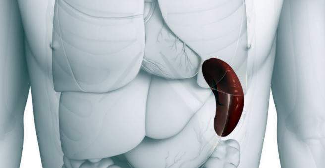
A splenic rupture is a rupture through the spleen tissue or capsule. The cause of this are mainly blunt abdominal injuries. A splenic tear is an acute emergency that needs to be treated as quickly as possible. Read all about symptoms, diagnostics and therapy of the splenic rupture!

Milz tear: description
The spleen is the most frequently injured organ in a belly trauma. In 30 to 50 percent of abdominal trauma, the spleen is affected.
One distinguishes a one-time and two-time splenic rupture. While in a single-stage splenic tear the capsule and spleen tissue break at the same time, in a two-period splenic rupture, only the spleen tissue is initially damaged and the dreaded tear of the capsule occurs hours to weeks later.
Spleen: anatomy and function
The spleen is an organ in the left upper abdomen under the ribs near stomach, kidney and intestine. It is surrounded by a connective tissue capsule. Some people have a small extra spleen (minor spleen). The main task of the spleen is the “filtration” of the blood for immunological monitoring and the degradation of red blood cells and platelets. Because of these tasks, the spleen’s special blood vessels are always filled with blood.
Splenesis: symptoms
One of the most common signs of splenomegaly is a (pressure) pain in the left upper abdomen, which may also radiate into the left flank and the left shoulder (sweeping sign). Depending on the extent of the splenic rupture, the abdominal wall can be very sensitive to pain and hard. In severe cases, respiratory distress, low blood pressure, high heart rate shock, collapse, and cardiovascular arrest may occur.
If the splenic rupture is due to trauma, the bump marks in the left upper abdomen or ruptures of the ribs in this area are noticeable. In a traffic accident, a bruise along the seat belt in the left upper abdomen may indicate severe trauma to the spleen.
In the case of a so-called two-time splitting of the spleen, the initial pain may initially fade away to return (intensified) after a pause (“silent interval”).
Milzriss: causes and risk factors
The most common cause of a splenic rupture is a blunt abdominal injury, despite protection from the ribs. Such a splitting usually occurs in traffic accidents. But even with falls events from high altitude and sports such as skiing, snowboarding and mountain biking can lead to a splenic rupture. A common accident mechanism is, for example, the fall over the bicycle handlebars.
In children, the ribs are even softer and the abdominal muscles weaker than in adults, which is an increased risk of injury. In particular, the seat belt in the car can cause a splitting in a traffic accident by the fixed train. In children should be thought in appropriate suspicion but also a child maltreatment as the cause of splenic rupture.
An important risk factor for increased bleeding in a splenic tear is a blood-thinning treatment such as that used in certain cardiac arrhythmias, thromboses or mechanical heart valve replacement.
A non-injury-induced splenic rupture can have various causes. In most cases, the underlying disease initially leads to enlargement of the spleen, which increases the tension of the spleen capsule. This in turn increases the risk of a spontaneous splenic rupture.
infections
The Pfeiffer’s glandular fever (also called infectious mononucleosis) is a virally induced, flu-like disease that can lead in rare cases by spleen enlargement to a spontaneous splitting of the spleen. To prevent this, patients are advised to take a sports break for four to six weeks.
Other infections with a risk of splenic rupture include malaria and typhoid fever.
inflammation
Even heavy or lengthy inflammations can lead to spleen enlargement due to the upregulation of the immune system. These include chronic pancreatitis, liver inflammation, autoimmune diseases and amyloidosis. Amyloidoses are usually deposits of abnormally altered proteins that affect the whole body.
tumor
The spleen can be affected by cancer. In most cases, these are tumors of the hematopoietic system such as lymphoma (non-Hodgkin’s lymphoma, etc.) or leukemia (acute or chronic myeloid leukemia, etc.). Less common are vascular tumors (angiosarcomas) or secondary tumors of tumors (metastases).
Diseases of the blood
The spleen is responsible for the breakdown of blood cells. Therefore, it is involved in most diseases of the blood. If, for example, platelets in the context of the Werlhof disease (ITP) are increasingly marked and attacked by their own immune system, the spleen ablates them. Due to the increased “burden” of the spleen, it increases. This is similar with the so-called hemolytic anemias – forms of anemia, which are based on an increased destruction of red blood cells.
Innate and structural causes
Also disturbances of the spleen structure, which lead to a blood back pressure, for example, can cause splenomegaly and splenic rupture. These often include congenital tumors of the blood vessels (hemangiomas) or cysts of the spleen. Such tumors can cause massive bleeding and thus a splenic rupture.
If the spleen is too loosely anchored in the surrounding tissue, it can easily lead to splitting of the spleen as well as distortions and disturbances in the blood flow in the vessels. The spleen may also be affected by pregnancy or blood backlog due to liver disease (portal hypertension).
Operations on the stomach
In abdominal surgery there is a risk of injury to the spleen or its vessels. How high the risk of a splenic rupture during surgery depends on many factors. These include above all the individual anatomy, the proximity of the operating area to the spleen and the experience of the surgeon. In the event of a severe spleen injury during surgery, it may be necessary to remove the spleen immediately if the surgeon does not appear to be conservative in treatment.
Splitting: examinations and diagnosis
Specialists in splenic rupture are surgeons. The suspicion of a splenic tear is an emergency situation and should be investigated as soon as possible. Therefore, the rescue service should be alerted especially for injuries that affect the left upper abdomen. Heart rate and blood pressure in the acute phase should be continuously monitored using monitor monitoring to assess circulatory stability. The doctor will ask questions such as:
- Did you recently have a stomach injury (about a beat)?
- Do you feel an abdominal pain?
- Did you have a fever or are you feeling sick?
- Do you have pre-existing conditions?
- Which medications do you take?
After that, the doctor will carefully examine the patient, especially the heart, lungs and abdomen. Following an accident, pay attention to bounce and belt marks and other external signs of injury. Typical for a splenic rupture is a pain in the left shoulder in addition to the abdominal pain. In addition, in some cases pain may be triggered by pressure on the left lateral neck (Sägesser sign).
Ultrasonic
The ultrasound examination is the quickest and easiest way to rule out acute bleeding in the abdominal cavity (FAST-Sono). If in doubt, the ultrasound should be repeated regularly. Contrast administration during ultrasound examination can improve the accuracy of the diagnosis.
Computed Tomography
The best method for detecting a splenic ridge is computed tomography (CT) with contrast medium. A particular bright area outside of vessels may indicate bleeding and spleen disorders. An X-ray or CT scan of the ribcage should rule out a bony injury to the ribs in an appropriate (accident) story.
laboratory tests
Any suspicion of a splenic rupture should be taken for analysis. In the laboratory, among other things, parameters for estimating blood loss through splenic rupture (hemoglobin, hematocrit, blood count) can be determined. Through repeated blood withdrawals, at the beginning also hourly, also changes of these values can be used as course parameters.
Milzriss: severity
Based on the exact diagnosis, the severity of the splenic rupture can be estimated. This helps planning the treatment planning and estimating the prognosis. In the classification according to Buntain and Gold one distinguishes four degrees of splitting:
- Local tear of the capsule or bruise under the capsule
- Capsule or tissue rips (except large spleen vessels)
- Deep cracks, which also affect the large spleen vessels
- Complete splitting of the spleen
There are a number of other systems for assessing splenic injury, some of which also involve the accurate assessment of the CT image.
Splitting: treatment
If the spleen is torn, in the emergency situation the stabilization of the circulation through fluid administration and medication via a venous access is at first in the foreground. It may also be necessary to have a blood transfusion depending on the blood tests.
After the initial examination, the decision must be made as to whether an emergency operation is necessary or whether one waits for the time being and carefully supervises the patient medically. The more severe the injury, the sooner an immediate surgery is sought. Even if the doctor suspects a hemorrhage in the abdomen and the circulation is unstable, an emergency operation is performed.
The right and quick choice of a spleen rupture is a crucial step and should be taken by an experienced doctor. Nowadays, splenic rupture can be treated in circulatory stable patients to a third degree spleen injury without surgery.
Conservative treatment
If no (immediate) surgery is performed, the patient should definitely be monitored in the hospital, including the intensive care unit if necessary. Especially in the first 24 hours after admission a strict bed rest should be kept. Circulatory parameters (such as blood pressure and heart rate) are monitored using a monitor. In addition, depending on the severity of the injury, close blood collection and ultrasound checks should be performed. In many cases, the risk of a severe course drops significantly after 72 hours.
surgery
There are a variety of different techniques for operating a splenic rupture. The most radical measure is the removal of the whole spleen (splenectomy). The condition without spleen is called asplenia. Splenectomy due to a splenic tear is nowadays almost exclusively performed with unstable circulation, evidence of bleeding in the abdominal cavity, and other signs of a severe splenic fracture.
In other cases, it is possible to remove only part of the spleen (spleen resection) or to aim for a vascular occlusion in the affected area. The latter is done with the help of suture, glue, electrical or other hemostasis (such as heat coagulation) or by packing the spleen into a resorbable net. Thereafter, the patient must be monitored in the hospital for approximately ten days if bleeding occurs despite the procedure.
It is also possible today to close individual (spleen) vessels with a catheter inserted into the inguinal veins (embolization) in order to stop active bleeding. However, this is associated with the risk that a spleen area supplied by the closed vessel dies.
Complications of the operation
Decisive for the postoperative course are regular follow-up checks. Abdominal pain up to several weeks after a stomach operation is possible.
Although the spleen is not an essential organ, the complete removal of the spleen entails some risks. Because it helps to defend the body against pathogens, those affected are at increased risk of infection after splenectomy.
In addition, any surgery in the abdomen carries general risks. In addition to injury to other abdominal organs (bowel, etc.), bleeding, infection and allergic reactions, pancreatitis and portal vein thrombosis may occur after spleen surgery.
In the so-called interventional procedures with catheters through the inguinal vessels, there is above all the risk of an unintentional vascular injury with bleeding or the formation of a vessel erosion (aneurysm).
Other possible complications include pseudocysts, abscesses, and arteriovenous shunts (abnormal connections between an artery and a vein).
asplenia
A life without spleen is associated with an increased risk of infection. For this reason, people without spleen must be regularly vaccinated (especially against pneumococci, meningococci and hemophilus influenzae). In case of fever sufferers should go to the doctor quickly. On the other hand, long-term prophylaxis with antibiotics against bacterial infections is only considered in children.
In asplenia, the so-called “OPSI” (overwhelming post-splenectomy infection), which leads to severe sepsis, is particularly feared. Especially toddlers and babies without spleen are at great risk for such a severe infection. This form of sepsis usually occurs in the first years after a splenectomy. However, an “OPSI” is still possible decades later. The main causes of this infection are pneumococci, hemophilus, meningococci, staphylococci and also E. coli strains.
In addition, the breakdown of platelets (platelets) by the spleen is eliminated so that in the first three months after the spleen, the number of platelets increases until the body has adjusted. Treatment with acetylsalicylic acid (and also heparin) to reduce the risk of thrombosis due to the high number of platelets is therefore recommended.
Milzriss: Disease course and prognosis
Decisive for the prognosis in a splenic rupture is the early diagnosis and the right therapy. Today, the vast majority of splenic tears can be treated without surgery. In addition, the rate of mitotic interventions or interventions increases, also thanks to the possibility of embolization. Especially in children, more than 90 percent of spleen injuries can be treated without surgery.
If only part of the spleen is removed, the remnant spleen can “regrow” and thus perform its full function again.
In up to four percent of patients whose spleen has been removed, there is “high blood mortality” (sepsis).
Life-threatening consequences of an injury usually appear within the first 24 to 72 hours after the injury. About 80 percent of patients with ruptured spleen are considered cured within seven to eleven weeks. Until then, the physical activity should be reduced so as not to pollute the spleen.