The hemangioma (blood sponge, blood sponge) is a benign vascular tumor in newborns and infants. It shows up as a red skin patch, which can be sublime. A hemangioma can occur on all parts of the body. In some cases, it will disappear by itself. Otherwise, it can be treated by various therapies. Read all important information about the hemangioma here.
Hemangioma: description
A hemangioma is a benign (benign) tumor of the vessels (angiodysplasia), which may be at different depths in the skin. Colloquially, it is also referred to as Blutschwamm or Blutschwämmchen. Hemangiomas do not form metastases, but they can press through their growth against organs and cause symptoms.
Hemangioma: types and frequency
A blood sponge occurs in infants and is either present from birth (congenital hemangioma) or develops in the first few weeks of life (infantile hemangioma). The latter is more common than the congenital variant.
Girls are about three times more likely to be affected by a sponge than boys. About five percent of premature infants and over 20 percent of preterm infants have an infantile hemangioma.
Lymphangioma is similar to hemangioma. The difference is that a lymphangioma arises from lymphatic vessels.
Hemangioma: symptoms
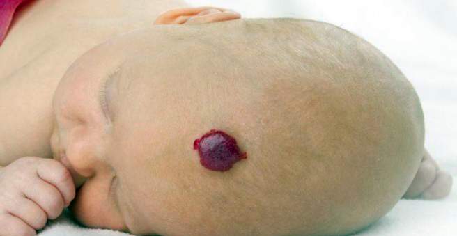
Hemangiomas are found mainly in the skin. Parents perceive them as red-blue dots, spots or knots. Sponges may be flat or sublime. The infantile hemangioma develops in the first four weeks of life. Then it can grow until about the ninth month of life.
Segmental hemangiomas are areally found in certain areas of the face and lower back, respectively. They often occur along with other malformations, such as brain hemangiomas or spinal malformations (such as spina bifida).
Hemangioma: causes and risk factors
The exact mechanisms that lead to a sponge are not yet fully understood. It is noticeable that hemangiomas are more common in certain families. This indicates an inheritable component in the formation of Blutschwämmchen out.
If someone has more than ten sponges, it is called hemangiomatosis. Hemangiomas are also common in internal organs such as the liver, so further investigations are necessary. Genetic syndromes such as the Kasabach-Merritt syndrome may also be associated with increased hemangiomas. In addition to the formation of large blood sponges on the extremities, there is a drop in the platelet count (thrombocytopenia).
Hemangioma: examinations and diagnosis
If you notice a red spot on your child’s skin, visit his pediatrician with him. First of all, this will ask you in detail about the history of the child’s illness (anamnesis). He will ask you the following questions:
- When did you notice the skin change for the first time?
- Has the size or color changed since then?
- Was there already a hemangioma in your family?
The doctor then examines your child. He takes a close look at the area of the skin that struck you. The rest of the skin is also searched for changes. In addition, the heart and lungs stop and check the movements of the child. This way, any associated malformations can be detected.
Further investigations
The history and clinical examination are crucial to diagnose hemangioma. Then the sponge should be photo-documented to detect changes over time. In addition, additional examinations are necessary in some cases. This includes an ultrasound examination (sonography). Hemangiomas can be detected in the abdomen such as in the liver. With magnetic resonance imaging (MRI), hemangiomas can be diagnosed in the brain.
Hemangioma: treatment
There are several ways in which a hemangioma can be treated. When choosing the method, it depends mainly on where the sponge is and how big it is. Rapid therapy should be used if the function of the tumor limits organs, such as the eye, ear, nose, mouth, feet or hands.
Also for cosmetic (facial) or nursing (genital) reasons, rapid therapy should be initiated.
Cold and laser therapy
Small, flat hemangiomas are usually treated with cryotherapy (cryotherapy). The Blutschwämmchen are thus iced. Prior to treatment, the affected skin area should be anesthetized with a local anesthetic patch. The same applies to a laser therapy. This can be used for very small Blutschwämmchen.
drugs
For larger or multiple hemangiomas, therapy with the drug propranolol is often preferred. Propranolol is a beta-blocker – a vasoconstrictor medication normally used in cardiovascular diseases. It is not officially approved for hemangioma therapy, so it is used off-label because it has been discovered by chance that it works quite well against blood sponges.
Propranolol should be given under hospital supervision. The dose is initially very low and then increased slowly, otherwise it can lead to cardiovascular disorders. In addition, the patient’s heart must be examined by means of electrocardiography (ECG) and cardiac ultrasound (echocardiography) before beginning therapy. Thus, any heart disease can be detected, which speak against a therapy with propranolol.
In the past, blood sponges have also been treated with glucocorticoids (cortisone) or chemotherapeutics, but this is now considered obsolete.
surgery
An operation is indicated for a hemangioma if, after completion of the other therapies, remnants of the blood sponge remain or scarring has developed. If rapid functional growth of the blood sponge threatens acute functional restrictions, such as obstructed nasal breathing, surgery should also be performed.
Hemangioma: disease course and prognosis
The prognosis is good. Infantile hemangiomas usually regress on their own, between the end of the first year of life and the ninth year of life. Often, no residues remain recognizable. In the case of very large sponges, on the other hand:
- scar
- swelling
- pigment changes
- skin thinning
A congenital hemangioma remains more common than an infantile one. With the right therapy you can completely eliminate it.