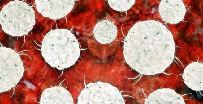
Hodgkin’s disease (Hodgkin’s disease, lymphogranulomatosis) is a malignant tumor of the lymphatic system (lymphoma). The disease is caused by degenerated white blood cells (B lymphocytes) in the bone marrow. Hodgkin’s disease is a rare cancer that primarily affects people around the age of 30 or 60 years. Men are more likely to get sick than women. Swollen, painless lymph nodes are typical symptoms of Hodgkin’s disease. Which therapy is used depends on the stage. There is also a chance of recovery in later stages of the disease. Read all important information about Hodgkin’s Disease here.

Hodgkin’s disease – description
Hodgkin’s disease is a rare cancer that affects the lymphatic system. The task of the Lyphsystems is, among other things, the formation and maturation of white blood cells (lymph cells = lymphocytes). The disease is named after the so-called Hodgkin cells. These cells are not found in healthy humans and can only be detected in tissue samples (biopsies) of patients with Hodgkin’s disease.
In addition to the lymphatic vessels, the lymphatic system also includes the lymphatic organs: bone marrow, thymus, spleen, lymph nodes and tonsils. The lymphatic vessels transport the lymph fluid out of the tissue back into the venous system. Between the individual sections of the lymphatic vessels are the lymph nodes, which act as filters. In them also degenerated lymphatic cells collect. Therefore, the lymph nodes are affected in a lymphoma at an early stage of the disease.
There are two groups of lymphoid cells: B lymphocytes and T lymphocytes. Both types are formed in the bone marrow and mature in various lymphoid organs. Normal B lymphocytes produce antibodies that help fight viruses and bacteria. Degenerate the B lymphocytes, a Hodgkin’s disease develops. Among other things, changes in the nucleus cause the cells to divide faster than healthy cells. The diseased cells first collect in Hodgkin’s lymph nodes, where they multiply and cause swelling.
The disease, also known as “Hodgkin’s Lymphoma”, accounts for only a relatively small group of all lymphomas, accounting for about 15 percent. Although Hodgkin’s disease can occur at any age, people between the ages of 25 and 30 and between 50 and 70 are particularly likely to be affected. Men are more likely to develop Hodgkin’s disease than women.
Histological forms of Hodgkin’s disease
There are four types of Hodgkin’s disease. Physicians divide them according to the type of affected tissue and the cell types that have settled there.
How good the prognosis of a patient with Hodgkin’s disease is depends on the type of disease. Physicians distinguish:
- Hodgkin’s disease of nodular-sclerosing type (60 percent) has good chances of recovery.
- Hodgkin’s disease of mixed type (30 percent) has a slightly worse prognosis than the nodular-sclerosing type.
- Lymphocytic-type Hodgkin’s disease (five percent) has good chances of recovery.
- Hodgkin’s disease of the lymphocyte-poor type (less than one percent) has an unfavorable prognosis
All four histological types show specific cells that are characteristic of Hodgkin’s disease. The so-called Sternberg-Reed cells are particularly large and have several cell nuclei. They are caused by the fusion of several degenerated B lymphocytes (Hodgkin cells).
Hodgkin’s disease – symptoms
Typical of Hodgkin’s disease painless, swollen lymph nodes in the neck area. Other lymph nodes may also be affected, for example under the armpits or the groin, but also in the thorax, where the doctor can not feel them, but only sees them on an X-ray. The lymph nodes are permanently enlarged in a Hodgkin lymphoma, or they continue to increase in the course of the disease on. Another typical feature is that they are difficult to move under the skin.
Enlarged lymph nodes can also have harmless causes. Thus, the lymph nodes also swell in the context of infections. However, these nodes react somewhat painfully to pressure, for example during palpation. In addition, they can be moved well under the skin. As a rule, the swelling recedes appreciably already a short time after the infection.
B symptoms Hodgkin’s disease
In addition to the main feature of Hodgkin’s disease, the swollen lymph nodes, more characteristic cancer symptoms occur. Doctors call her “B symptoms“. These are three typical signs that appear not only in Hodgkin’s disease, but also in other cancers and chronic infections:
- weight loss: A weight loss of more than ten percent of body weight in the previous six months.
- night sweats: Excessive sweating at night causes those affected to wake up “soaking wet” and have to move into bed and change their clothes.
- fever: Unexplained fever above 38 ° C
The symptoms of B in Hodgkin’s lymphoma often go in spurts. This means that the symptoms, aside from weight loss, can completely disappear for a few days and then recur.
As part of a Hodgkin’s disease, many sufferers from a sense of fatigue, Patients with Hodgkin’s disease are quickly exhausted and less efficient than they used to be. In addition, many feel tired all the time, even though they sleep well.
Ten to 25 percent of patients with Hodgkin’s disease also develop a generalized itching, The exact cause of this phenomenon is not yet known. It is believed that the degenerated blood cells release chemical substances near the sensitive skin nerves that trigger the itching.
In the advanced stage of Hodgkin’s disease, various other organs such as the liver, spleen, nervous system and bone marrow can also be affected by cancer cells. As blood builds up in the bone marrow, Hodgkin’s disease can affect the formation of all blood cells (pancytopenia). Possible consequences are anemia, a lack of platelets (thrombocytopenia) and a lack of functional white immune cells (leucocytes). The lack of blood formation causes a general weakness. In addition, patients are more likely to bleed and contract infections more quickly.
A rare, but very specific symptom in Hodgkin’s disease is the so-called alcohol pain, The affected lymph node region hurts after drinking alcohol. Even relatively small amounts of alcohol (for example, a glass of beer) are enough to trigger the pain. The exact mechanism behind the phenomenon is not yet known.
Especially in Hodgkin’s lymphoma, the so-called Pel-Ebstein fever occurs. Fever-like intervals of about three to seven days alternate with fever-free intervals.
Hodgkin’s Disease – Causes and Risk Factors
The exact causes of Hodgkin’s disease have not yet been clearly identified. Among other things, Hodgkin’s disease is linked to viral diseases. The Epstein-Barr virus (EBV), which causes the Pfeiffer’s glandular fever (mononucleosis) plays, according to the current state of knowledge, an important role in the development of Hodgkin’s disease. Thus, the virus can be detected in the lymphoma cells in 50 percent of patients suffering from Hodgkin’s disease.
Scientists also suspect a link between Hodgkin’s disease and HIV infection. The viruses are said to increase the susceptibility of the cells to changes, making it easier to cell degeneration and thus to Hodgkin’s disease.
Prior chemotherapy or radiation also appears to significantly increase the risk of Hodgkin’s disease. The toxic substances can alter the genetic material in the cell nucleus so that the degenerated Hodgkin’s disease cells develop. Since various substances in tobacco smoke can also damage the genome of cells, there may also be a connection between Hodgkin’s disease and smoking.
Hodgkin’s Disease – Examinations and Diagnosis
The right contact for suspected Hodgkin’s disease is your family doctor or a specialist in internal medicine and oncology. Already by describing the symptoms, the doctor receives important information about your state of health. There are many other diseases that can trigger lymph node swelling. These include various viral and bacterial infections and diseases of the immune system. If you suspect Hodgkin’s disease, the doctor will ask you about your history (anamnesis). Possible questions can be:
- Did you notice swelling on the neck?
- Have you recently been waking up in a sweaty night?
- Have you lost body weight in the past six months without your lifestyle changing?
- Have you had a fever lately?
- Has drinking alcohol caused pain recently?
Following the first conversation is usually a physical examination, The doctor is looking for swollen lymph nodes. Since enlarged lymph nodes are not specific for Hodgkin’s disease and may have other causes, a detailed laboratory diagnosis is then carried out. In addition, the doctor will scan liver and spleen, which may also be enlarged in Hodgkin’s disease.
blood test
With a blood test, the number of white blood cells can be determined. The typical Hodgkin’s disease blood count often shows an increased blood cell lowering rate (ESR). It measures the rate at which the solid blood components in a tube fall. How fast that happens depends on the surface of the blood cells. In inflammation or tumors, more proteins are found in the blood and on the cell surfaces. As a result, the blood cells adhere to each other, forming larger heavier units that sink faster.
In advanced stages, Hodgkin’s disease can also affect bone marrow. In the blood picture this manifests itself in the form of anemia (anemia) and a lack of platelets (thrombocytopenia) and white defense cells (leukocytes). Also typical for the blood picture of Hodgkin’s lymphoma is an excessive proliferation of certain white blood cells, the eosinophilic granulocytes (eosinophilia). It occurs in about one third of the cases.
Tissue sample bone marrow & lymph nodes
If there is a suspicion of Hodgkin’s disease, a complete lymph node is removed (lymph node extirpation) to confirm the diagnosis and examined under the microscope. In Hodgkin’s disease, B lymphocytes degenerated into so-called Hodgkin cells, which regularly have a nucleus. When several Hodgkin cells merge, a Sternberg reed cell is created. Under the microscope, one then recognizes very large cells with several cell nuclei. Their appearance is considered evidence of the presence of Hodgkin’s disease.
Another diagnostic possibility provide tissue samples from the bone marrow (biopsy). The doctor pulls out the sample with a puncture needle from the marrow of the iliac crest. Such a bone marrow tissue sample may also contain degenerate cells, giving evidence of Hodgkin’s disease.
Imaging procedures
Imaging examinations such as x-rays, ultrasound, computed tomography (CT) or skeletal scintigraphy help to determine the stage of the disease and to detect possible secondary tumors (metastases) in other organs.
Hodgkin’s disease – staging (to Ann-Arbor)
Hodgkin’s disease is divided into four stages, depending on how much it has spread throughout the body. The more lymph node regions are affected, the more advanced the disease and the worse the prognosis. Basically, Hodgkin’s disease is a cancer that can be cured at any stage.
|
stage |
Infestation of the lymph nodes |
|
I |
Infestation of only one lymph node region |
|
II |
Infestation only on one Side of the diaphragm: (Lymph nodes either in the chest or in the abdominal area affected); |
|
III |
Infested both Sides of the diaphragm: (lymph nodes both in the thorax and in the abdomen), |
|
IV |
Infestation of one or more extralymphatic Organs (brain, bones) independent of the pattern of involvement of the lymph nodes. |
Hodgkin’s disease – treatment
The goal of the treatment is a complete cure in each of the four stages. The therapy of Hodgkin’s disease is based on two forms of therapy: chemotherapy and radiotherapy. In early stages, when Hodgkin’s disease is still confined locally to a region, radiation is at the forefront. In advanced stages, intensified chemotherapy is needed for healing.
In the Stages I and II Chemotherapy is carried out according to the so-called ABVD scheme in two cycles. The patients receive a combination of four chemotherapeutic agents – Adriamycin, Bleomycin,Vinblastin,Dacarbazin. There is also a radiation therapy
In the Stage III and IV the attending physician sets the BEACOPP scheme (Bleomycin, etoposid, Adriamycin, Cyclophosphamid, Oncovine (vincristine), Procarbazin, Prednison). With positron emission tomography (PET), he can search for remaining remnants of the tumor after chemotherapy. E is optionally again selectively irradiated.
For irradiation, the so-called involved-field technique is used. In doing so, the doctor attempts to irradiate only the area affected by tumor tissue and to protect the adjacent healthy tissue.
Prognostically unfavorable risk factors are also taken into account in treatment planning. The unfavorable prognostic factors include: a high blood cell lowering rate, a large tumor in the middle of the chest cavity (mediastinum) and other affected organs (stage 4).
Hodgkin’s Disease – Disease Course and Prognosis
Fortunately, the Hodgkin’s disease chances are very good. In the last 30 years, the therapeutic options and thus the prognosis have improved considerably. Nevertheless, it is important to recognize the disease as early as possible and treat it accordingly. The five-year survival rate after therapy is 80 to 90 percent. This means that 80 to 90 percent of those affected are still alive five years after diagnosis. In children, the five-year survival rate of Hodgkin’s disease is even over 90 percent. The sooner a Hodgkin’s disease is detected, the better the chances of recovery. Even at an advanced stage, however, healing is still possible. Also, Hodgkin’s disease recurrence, when the cancer returns, good long-term treatment outcomes can be achieved. However, years or decades after the Hodgkin’s disease Treatment due to chemo and radiotherapy to develop another cancer (thyroid cancer, breast cancer, leukemia, etc.).