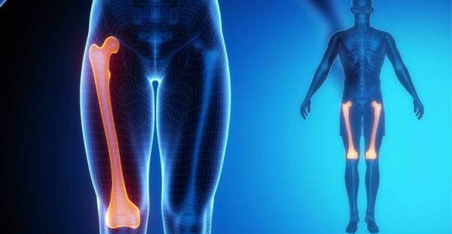
A femur fracture (thigh fracture) occurs when strong forces act on the bone, such as in an accident. Depending on the location of the fracture, a variety of femoral fractures are distinguished. In all cases, typical symptoms are severe pain, swelling and a malposition of the leg that can no longer be stressed. The treatment usually consists of surgery. Everything important about the femur fracture can be found here.

Femoral fracture: description
In a femoral fracture (femur fracture) the longest bone of the body is broken. Such an injury rarely occurs alone, but usually in the context of trauma such as serious car accidents.
The femur (femur) consists of a long shaft and a short neck, which also carries the ball of the hip joint. In the area of the shaft, the femur is very stable. The greater trochanter, a bone protrusion between the femoral neck and the shaft, serves as a muscle attachment site. The lesser trochanter is a small bony prominence on the inside of the femur.
A femoral fracture is subdivided into:
- Femoral neck fracture
- Pertrochanteric femur fracture
- Subtrochanteric femur fracture
- Hip-joint-proximal femur fracture (proximal femur fracture)
- femoral shaft fracture
- Knee joint-proximal femur fracture (distal femur fracture)
- Periprosthetic femur fracture
In the following, all types of fractures are considered in more detail – with the exception of the femoral neck fracture. This is dealt with in more detail in the article .
femoral shaft fracture
The thighbone is surrounded by a strong soft-tissue mantle, which consists of the quadriceps muscle at the front and the ischiocrural musculature at the back. On the inside is the adductor group, another muscle group. Depending on the location of the thigh fracture, the bone elements are displaced by the musculature in a certain direction.
Hip-proximal femoral fracture
In 70 percent of all thigh fractures is a proximal femur fracture. The fracture gap is located further up the shaft near the hip joint. In this form of femoral fracture, the upper bone fragment is rotated outward by the musculature.
Knee joint (distal) femur fracture
The distal femur fracture (also known as supracondylar femur fracture) is localized to the shaft near the knee joint. The upper bone fragment is pulled on the inside and the lower fragment pushed backwards.
Pertrochanteric and subtrochanteric femoral fracture
The pertrochanteric fracture of the femur is a fracture of the femur near the hip, where the fracture line passes through the greater and minor trochanters. It is a typical injury in the elderly with osteoporosis. This form of femoral fracture occurs at least as often as the femoral neck fracture and accounts for approximately 40 to 45 percent of proximal femoral fractures.
The so-called subtrochanteric femoral fracture is a rupture below the trochanteric femoral shaft, and has approximately the same characteristics as a pertrochanteric fracture of the femur.
Periprosthetic femur fracture
A periprosthetic femur fracture occurs when the femur is anchored in a prosthesis, such as a hip or knee prosthesis. Because there are more and more people with such prostheses, the incidence of periprosthetic thigh fractures is also increasing.
Femoral fracture: symptoms
A femoral fracture is very painful. The affected leg can not be loaded, swells and shows a malposition. Frequently, an open fracture develops – the skin is injured by bone splinters.
The immediate measure at the scene of the accident is that the leg is stored and shaved as painlessly as possible. In the case of an open fracture of the femur, it is best to cover the wound with sterile dressings until the patient has arrived at the hospital.
A femoral fracture can cause large bleeding, potentially causing a circulatory shock. Symptoms of this are a cold-sweaty skin with a pale grayish color. Regardless of the ambient temperature, those affected shiver and shiver, their hands and feet are cold.
femoral shaft fracture
In a femoral shaft fracture, the leg appears shortened, and there is a significant malposition of the thigh visible. The patient is unable to bend the knee or lift the lower leg. A femoral shaft fracture is very painful. Even if only an isolated femoral stem fracture occurs, the patient may lose up to one and a half or two liters of blood.
Hip-proximal femoral fracture
In the proximal femur fracture, the leg appears shortened and rotated outward. Concerned also describe a compression pain and pain in the groin.
Knee joint (distal) femur fracture
Distinct fracture signs in a distal femur fracture are a bruise and swelling and possibly a fur position of the leg. The knee can not be moved. There is also a lot of pain.
Pertrochanteric and subtrochanteric femoral fracture
A typical symptom of a pertrochanteric femur fracture is a shortened and outwardly rotated leg. The person is uncertain when walking and standing. The leg can not be moved because of the severe pain. Sometimes you see a bruise or a bruise.
The subtrochanteric femoral fracture shows the same symptoms as the pertrochanteric fracture.
Periprosthetic femur fracture
Depending on the location of the fracture, a periprosthetic femur fracture may show symptoms similar to a normal femoral fracture. The rupture may occur around the greater trochanter, the shaft, and near the knee joint.
Femoral fracture: causes and risk factors
A femoral fracture occurs when strong forces act on the bone. Traffic accidents are common causes of a thigh fracture. This usually affects younger people. In the elderly, the femoral fracture usually occurs near the knee joint or femoral neck. Osteoporosis, in which the bone is decalcified, plays a major role in this. In contrast to the femur fracture, the femoral neck fracture occurs even in mild falls.
femoral shaft fracture
The thigh (femur) is the strongest bone of the extremities. Except for bone disease (including osteoporosis), a significant amount of trauma is necessary for it to break. The breakage can be a simple transverse, oblique or piece breakage. Even a floor or fragmentary fracture is possible, namely with a force on the entire thigh. Indirect violence in traffic accidents and falls from great heights may result in breakage with rotary wedges or bending wedges. Wound and explosion injuries cause defect fractures. About 20 percent of people with femoral shaft fracture have a polytrauma, ie simultaneous injuries in several body regions.
Hip-proximal femoral fracture
In 70 percent of all thigh fractures is a proximal femur fracture. It is a typical fracture of the old man. Accident is usually the domestic fall.
Knee joint (distal) femur fracture
Accident mechanism in the distal femur fracture is often a Rasanztrauma (Hochrasanzrauma) – a lot of kinetic energy (kinetic energy) acts on the bone. The result is usually a larger rubble zone, which often involves joints, capsules and ligaments. But even old people with osteoporosis can suffer a distal femur fracture, which is usually a simple fracture.
Pertrochanteric femur fracture
A typical accident in the pertrochanteric femur fracture is the direct fall on the hip.
Periprosthetic femur fracture
The cause of a periprosthetic femur fracture is usually a fall or accident. Risk factors are:
- Diseases like osteoporosis
- wrong position of the shaft in the prosthesis
- incomplete cement coat
- Bone tissue that dissolves (osteolysis)
- relaxed prosthesis
- repeated joint replacement
Another reason could be so-called “stress shielding”. This means that the prosthesis takes over the function of the bone and therefore the bone degrades due to the lower load.
Femoral fracture: examinations and diagnosis
In the extreme case, a femur fracture can be life-threatening, so if you suspect such a fracture, you should immediately call the primary care service or your family doctor. The specialist for fractures is the doctor for orthopedics and trauma surgery. For the diagnosis, the doctor will ask exactly about the accident and your medical history. Possible questions could be:
- How did the accident happen?
- Was there a direct or indirect trauma?
- Where is the possible fracture?
- How do you describe the pain?
- Were there any previous injuries or previous damage?
- Have you previously had symptoms such as stress-related pain?
This is followed by a physical examination. The localization, the pain and the malposition of the leg are clear indications for a femoral fracture. In addition, the doctor will examine you for vascular and nervous system injuries by checking motor function, sensitivity, and blood flow in the leg. He will also look for typical concomitant injuries such as acetabulum fracture (fracture in the acetabulum), an additional femoral neck fracture or ligament injuries to the knee joint.
Apparative diagnostics
An X-ray confirms the diagnosis. The fracture can be accurately assessed if the entire thigh is x-rayed with its adjacent joints. It also takes pictures of the pelvis, hip joint and knees in two levels.
For debris or defect fractures, a comparison of the opposite side is usually made for further treatment planning. If vascular injuries are suspected, Doppler sonography or angiography (vascular X-ray) may be helpful.
Femoral fracture: treatment
Already at the scene of the accident, the leg should be splinted and carefully pulled out. Therapy in the hospital is usually to stabilize the leg surgically. For this purpose, you must set up the fracture anatomically accurate and restore the axis and rotation without loss of function.
femoral shaft fracture
A femoral shaft fracture is usually operated on. The technique usually used is the so-called locking nailing. Thus, the femur generally heals faster and can be charged earlier. In addition, only a few soft tissues are injured during the operation.
In patients with unstable paroxysm and an open, contaminated fracture, the femoral fracture should first be treated with a lateral “external fixator” – a support frame placed on the outside of the bone to immobilize it. For a femur fracture with severe soft tissue damage and concomitant thoracic trauma (injury to the chest and its organs) particles of the bone marrow can be washed with the blood into the lungs and there cause a so-called fat embolism: The alluvial fat clogged thereby the blood vessels of the lungs and thereby affect the oxygen supply. Only when the leg has stabilized, a further treatment may take place.
After the operation, the doctor tests the stability of the knee joint. This is important because cruciate ligament injuries are particularly common in younger patients with femur fracture due to high-rasping trauma.
Femoral shaft fracture in children
In newborns, infants and toddlers with a femoral shaft fracture physicians first try a conservative treatment. A closed fracture can be immobilized for about four weeks with a pelvic / leg cast or a hospital overheed extension (the leg is raised vertically). In rare cases, surgery is considered.
In school age, the operation is preferable to a femoral shaft fracture. A pelvic leg encounters difficulties in home care at this age. An extension is just as hard to carry out because of the long time in the hospital and the discomfort. Depending on the injury, it is primarily treated with the “external fixator” and in less complicated cases the elastically stable intramedullary nailing (ESIN) is performed.
Hip-proximal femoral fracture
The technique in the therapy of femoral fracture has developed in recent years. Meanwhile new implants are available for osteosynthesis. After the operation of a proximal femur fracture, the patient can usually start early with movements and quickly re-integrate into his usual environment.
Knee joint (distal) femur fracture
In a femoral fracture near the knee joint or involvement of the articular surface, it is important that the bone be accurately anatomically aligned again. Only this will give you a good functional result.
The conventional methods stabilize the fracture with angle plates and the dynamic condyle screw (DCS). In the meantime, recent developments have emerged: the so-called retrograde technique of intramedullary nail osteosynthesis and inserted plate systems have shown good results, whereby the screws are anchored in the plate with stable angles.
Pertrochanteric femur fracture
In the case of pertrochanteric femoral fracture, conservative treatment is virtually impossible because the fracture is very unstable. Therefore, surgery is performed – as well as in the subtrochanteric femur fracture. Dynamic screws anchored in the femoral head stabilize the fracture. They are then anchored with a plate (dynamic hip screw, DHS) or a nail (gamma nail, proximal femur nail, PFN) angle stable. A sliding mechanism applies pressure to the fracture as soon as the leg is loaded. This type of surgery is a minimally invasive surgical technique that protects the surrounding soft tissue. Even with a highly unstable pertrochanteric fracture of the femur, the leg can be fully loaded again after surgery.
Periprosthetic femur fracture
In the case of periprosthetic femoral fracture, the operation is preferable to conservative treatment. Depending on the fracture type, various operations such as replacement of the prosthesis, plate osteosynthesis or retrograde nailing are used.
Aftercare for femoral fractures
The post-treatment depends on how severe the injuries are and how stable the osteosynthesis is. After the operation, the leg is stored on a foam rail until the wound drainage is removed. Two days after the procedure, a passive movement therapy with the so-called CPM movement rail is started. Depending on the course of the femur fracture and the implant, the leg can be partially loaded slowly increasing again. How much stress is allowed depends on how much callus (new bone tissue) has formed. This is checked in the X-ray image. After about two years, the plates and screws are surgically removed.
Femoral fracture: disease course and prognosis
The prognosis of a femoral fracture depends on the type and extent of the fracture. Possible complications include:
- storage damage
- compartment syndrome
- Deep pelvic vein thrombosis (DVT)
- Infections, especially in the medullary cavity (especially with open femur fracture)
- pseudoarthrosis
- Angular deformity
- Rotation error (especially in the intramedullary nail osteosynthesis)
- leg shortening
- ARDS (acute respiratory distress syndrome): acute lung injury; possible complication of a severe trauma with shock
In the case of an uncomplicated course, the prognosis for a femoral fracture is generally good. Residual complaints such as swelling of the leg, numbness or weather sensitivity in the leg can persist for several months. But they usually disappear completely again.
femoral shaft fracture
The prognosis after treatment of femoral shaft fracture is very good. About 90 percent of cases heal out within three to four months without remaining. If the bone heals badly, an intramedullary nail osteosynthesis can be used to remove the locking pin and attach autologous (endogenous) cancellous bone (spongy tissue inside a bone). This stimulus can accelerate bone healing.
Hip-proximal femoral fracture
If the affected person can not fully re-stress his leg and be mobile, he will usually be in need of care.
Knee joint (distal) femur fracture
After the operation exercises can be started early. After about twelve weeks, the leg can usually be fully loaded again.
Pertrochanteric femur fracture
In a pertrochanteric femoral fracture The patient can use his leg completely right after the operation.