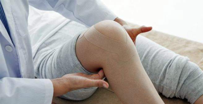
The Baker’s cyst is a fluid-filled protuberance in the popliteal fossa. It is caused by a weakness of the knee capsule and contains synovial fluid called synovial fluid. It is usually the result of another disease of the knee joint. It often causes no discomfort, but can press on vessels and nerves and then lead to complications. Read all about the development and treatment of the Baker’s cyst.

Baker’s cyst: description
The Baker cyst, sometimes called Baker cyst, is named after an English surgeon named William Baker. In the 19th century, he came across a cyst in the knee of some patients, more specifically in the popliteal fossa, and described this phenomenon first.
What is a cyst?
Cysts in medicine are cavities in the body that are not present under normal circumstances. They occur in various body tissues and organs (lungs, kidneys, liver, ovaries, etc.) and are either filled with fluid or, in the case of pulmonary cysts, with air. The Baker’s cyst is in many cases harmless and often goes unnoticed at first, but with increasing size usually problems arise.
How is the Baker’s cyst formed?
Like every joint, the knee is surrounded by a connective tissue-like covering, the joint capsule. On the one hand, it contributes to stabilization, on the other hand, its inner layer produces what is known as synovial fluid, or synovial fluid for short, a kind of “synovial fluid” that reduces friction at the articular surfaces. In addition, it supplies the articular cartilage with nutrients and contributes to the mechanical damping.
When the knee joint is damaged or inflamed, the body reacts by producing more synovial fluid. This increases the pressure in the joint capsule. If it is too big, the capsule can thinn out at a weak point and evacuate like a bag. Such a weakness lies in the knee capsule behind and then manifests itself as a cyst in the popliteal fossa. It typically develops on the inside of the popliteal fossa between the attachments of the gastrocnemius muscle (a calf muscle) and the semimembranosus muscle (a large posterior thigh muscle).
Who is affected by a Baker’s cyst?
Especially in the elderly, a Baker’s cyst often arises. Knee and other joint problems become more common with age and thus more likely to occur. Ultimately, however, a Baker’s cyst can occur at any age. However, children are much less affected than adults. At younger ages, a Baker’s cyst sometimes develops spontaneously, not as a result of knee damage. The reasons are not yet clear.
Baker’s cyst: symptoms
The bigger the Baker’s cyst, the more likely it will cause problems. In contrast, smaller cysts often remain without symptoms. The size of a baker’s cyst depends, on the one hand, on how long it has developed and, on the other, varies with the mechanical stress on the affected knee. The cyst and the load are related as follows: The body reacts to a heavy load on the already damaged joint with an increased inflammatory reaction and thus an increased formation of synovia.
Accordingly, a Baker’s cyst swells in addition, for example, by exercise or physical labor, in addition. Conversely, it will become smaller again as soon as the patient spares his knee for a few days. As long as you do not treat the underlying disease, the Baker’s cyst usually increases in volume and eventually causes symptoms. These may include:
- a palpable swelling in the popliteal fossa as soon as the Baker’s cyst has reached a certain size (from about 2 cm).
- the noticeable movement of fluid under the skin of the popliteal fossa. This phenomenon is called in medicine as fluctuation.
- initially an indefinite pressure on the back of the knee by the Baker’s cyst. The popliteal and upper calf can also become increasingly painful.
- Circulatory disorders and feelings of numbness to paralysis of the lower leg and foot.
The symptoms mentioned in the last point occur when the Baker’s cyst presses on vessels and nerves in the region of the popliteal fossa.
The burst Baker’s cyst
Complications arise especially when the Baker’s cyst ruptured, so tearing. This can happen if it gets too big and the pressure in relation to the wall thickness has reached a critical level. If the person then bends the knee, the thinned cyst wall can no longer withstand the pressure increase and tears.
Once the baker’s cyst has burst, the synovium will leak into the surrounding tissue, causing inflammation and additional pain. Following gravity, the leaked synovial fluid enters the lower leg muscles and in some cases even to the region around the ankle.
Due to the inflammation and swelling caused in the tissue pressure builds up there, which can not escape. Doctors then speak of a compartment syndrome. This presses on nerves and smallest blood vessels and can have serious consequences up to the loss of the lower leg, if not treated in time surgical treatment.
Baker’s cyst: cause and risk factors
An abnormally increased production of synovia in the knee joint capsule occurs especially when the knee joint is damaged. Be it due to wear, injuries or inflammation. The most common causes of a baker’s cyst are:
- Osteoarthritis: As people age, many people experience wear and tear on their joints. The knee joint is particularly stressed during physical activity.
- Meniscal damage: If one of the two cartilage discs breaks in the knee joint, for example as a result of an accident, more irritation will result from the irritation. This also affects younger people.
- Arthritis: Inflammation in the knee joint is often caused by rheumatoid diseases. In rarer cases but also bacteria are the trigger.
The two biggest risk factors for a Baker’s cyst are a higher age, as well as, among younger people, activities and sports that are associated with a high knee load. Occasionally, operations are the trigger of a Baker cyst. Knee-TEP surgery and cruciate ligament reconstructions would be examples.
Baker’s cyst: examination and diagnosis
Most patients seek advice from the orthopedist when the baker’s cyst is already larger and the first symptoms appear. Sometimes, however, it is also incidental when the knee is examined for other reasons. The doctor first asks for the medical history of the patient. He is particularly interested in whether problems with the knee joint have occurred in the past. The preliminary talk is followed by a physical examination, in which, in the case of a Baker’s cyst, a rounded, bulging, elastic swelling in the popliteal fossa can usually be felt.
But there are other reasons for such swellings such as tumors or thrombosis. Therefore, for the safe diagnosis of a fluid-filled cyst. Popliteal and calf muscles examined with an ultrasound machine. On the one hand, this allows the examiner to recognize the Baker’s cyst in the knee as a fluid-filled capsule bulge; on the other hand, any swellings in the calf muscles can be detected by spilled synovia.
Another method of investigation is magnetic resonance imaging (MRI), which can be used to detect fluid accumulation in the body. An MRI is more accurate than ultrasound examination and less dependent on the experience of the examiner. It also provides additional information on possible meniscal damage or joint wear. However, this examination method is also much more expensive and is therefore not used by default.
If the diagnosis “Baker’s cyst” is established, additional studies may be included to track down the causative disorder.
Baker’s cyst: treatment
Therapy differentiates between symptomatic and causal approaches. Symptomatic methods only relieve the symptoms caused by a baker’s cyst, while causal therapy addresses the root cause of the condition. Not every baker’s cyst needs to be treated. As long as she does not cause any problems, you can also wait.
Baker’s cyst: therapy with drugs
For the treatment of pain, the classic drugs from the field of nonsteroidal anti-inflammatory drugs (NSAIDs) are available. This group includes, for example, diclofenac and ibuprofen. In addition to relieving pain, they also counteract inflammation. Furthermore, there are so-called Cox-2 inhibitors, which act similar to the classic NSAIDs, but have fewer side effects on the gastrointestinal tract.
Cortisone is a naturally occurring hormone (cortisol) in the body, which has numerous effects, including a strong anti-inflammatory. Since it has significant side effects at high doses or long-term use, it must be used wisely. In the Baker’s cyst, the doctor can inject cortisone into the knee joint, so that the active substance there temporarily stops the inflammatory processes. But this should not happen more than three times a year.
Finally, there is the possibility of injecting hyaluronic acid into the joint. That sounds contradictory, because hyaluronic acid is indeed the main component of the Synovia, of which too much is actually present. However, hyaluronic acid improves the quality of cartilage tissue in the joint, the damage of which is often the cause of a baker’s cyst. Insofar, the use of this substance can achieve long-term positive effects.
Baker’s cyst: physiotherapy
Various physiotherapeutic measures help to alleviate the symptoms of a baker’s cyst. There are, for example, special strength and leg axis training or water training – methods that help the joints to strengthen the muscles around the knee joint and reduce the stimulation situation. However, depending on the underlying disease, it must always be clarified by a doctor whether physiotherapy is an option.
Baker’s cyst: puncture
It is possible to puncture a baker’s cyst and use a syringe to aspirate its liquid contents. This may be temporary relief for the patient, but it is very likely that the cyst soon fills up with Synovia and swells again.
Baker’s cyst: surgery and thermotherapy
To treat a baker’s cyst sustainably, the actual cause must be treated. For many patients, this sooner or later means an operation on the knee joint, for example to repair damage to the cartilage or the menisci. Such an intervention can be open or, minimally invasive, via a joint mirroring. The actual cyst is usually not removed, it goes back to elimination of the cause of alone. However, if rheumatoid arthritis is the cause, the surgeon removes the entire Baker’s cyst.
More recent techniques rely on bipolar current, which is applied to the cyst wall by means of electrodes, after having punctured the cyst contents. The resulting heat shrinks and clogs the cyst wall, preventing Synovia from overflowing.
Baker’s cyst: homeopathy
Homeopathic approaches to the treatment of a Baker’s cyst are used concomitantly with the aforementioned therapeutic methods. For example, after an operation, they should support the healing process. A commonly used remedy in homeopathy, which is also used in the Baker’s cyst, is Arnica C30.
The concept of Schüßler salts and their specific efficacy are controversial in science and not clearly established by studies.
Baker’s cyst: disease course and prognosis
The Baker’s cyst in many cases does not cause any problems as long as it is still smaller. Accordingly, therapeutic measures are only required if symptoms occur or if there is a risk of complications. Since the cyst in the knee is usually only the symptom of another disease and it also often increases creeping in size, sooner or later, the need for a therapy arises.
Although symptomatic treatments can alleviate the symptoms and delay surgery, spontaneous regression of the cyst is not expected. Many patients will only have the surgery Baker cyst permanently going on.