A pelvic fracture (pelvic fracture) is usually the result of severe force effects, such as may occur in the context of traffic accidents or falls. Patients often suffer from polytrauma, ie simultaneous injuries to various parts of the body. A pelvic fracture can be life-threatening due to the large blood loss. A mild pelvic fracture may be treated conservatively, while an unstable pelvic fracture usually requires surgical management. Everything important about the pelvic fracture can be found here.
Pelvic fracture: description
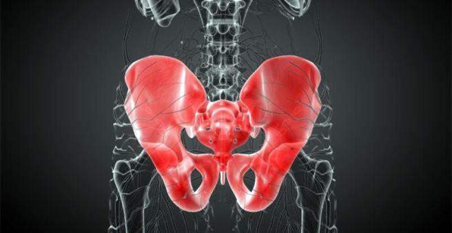
The pelvis is the connection between the spine and the legs and at the same time supports the intestines. It consists of several individual bones, which are firmly connected and form the pelvic ring. Basically, a pelvic fracture can occur at different sections of the pelvis.
Pelvic fracture: classification
Basically, a distinction is made in the pelvic fracture between injuries of the pelvic ring and the acetabulum. The Association for Osteosynthesis Issues (AO) divides the various pelvic ring injuries according to the stability of the pelvic ring. Roughly distinguishes a stable and an unstable pelvic fracture.
Stable pelvic fracture
The stable fracture of the pelvic fractures (type A pelvic fracture) usually refers to an isolated fracture of the pelvic margin, a front pelvic fracture, or a transverse fracture of the sacrum and coccyx. The pelvic ring is preserved throughout, so it is called a stable pelvic fracture.
Unstable pelvic fracture
An unstable pelvic fracture is a complete rupture involving the anterior and posterior pelvic ring. Physicians speak of type B, although the pelvis is vertically stable, but rotationally unstable. This concerns, for example, the symphyseal blasting “open-book injury”: The pubic symphysis is here apart, the two halves of the symphysis are opened like a book.
Furthermore, one speaks of a pelvic fracture type C, if it is a completely unstable pelvic fracture. The pelvis is ruptured by vertical gravitational forces and is unstable both vertically and rotationally.
Hüftpfannenfraktur
The hip socket fracture (acetabular fracture) is a joint fracture and therefore occupies a special position. It results from severe direct trauma when the femoral head is suddenly pushed against the acetabular cup, or indirectly, by passing the force over the femur. This can happen, for example, when the knee hits the dashboard in a rear-end collision. The acetabular fracture often occurs in combination with a hip dislocation. In some cases (15 percent), the peripheral nerve of the leg, the sciatic nerve (sciatic nerve), is also injured.
polytrauma
A pelvic fracture is a serious injury. In 60 percent of cases, patients with pelvic fractures also have injuries to other parts of the body (ie are polytraumatized). Especially the following injuries can occur in combination with a pelvic fracture:
- Fractures of the peripheral skeleton (in 69 percent of pelvic fracture patients)
- Traumatic brain injury (at 40 percent)
- Thorax injuries (at 36 percent)
- Injuries to the abdominal organs (at 25 percent)
- Spinal injury (at 15 percent)
- Urogenital injury (at 5 percent)
Pelvic fracture: symptoms
Pelvic fracture symptoms include swelling, pain, and possibly an unstable pelvic bone. In a simple, stable pelvic fracture, the complaints are usually less severe than an unstable pelvic fracture.
In addition, contusion marks or bruises may show on the dependent parts of the body such as the testicles, labia and perineum. In some cases, the pelvic fractures may cause the legs to have different lengths.
Unstable pelvic fractures often occur as part of multiple injuries (polytrauma). For example, bloody urine may indicate bladder injury, which is more common with pelvic fractures.
Frequently, the pelvic bones can easily be shifted against each other in the patients. In extreme cases, the pelvis works like a book (“open book”). Walking is no longer possible for a person with such an injury.
Pelvic hernia: causes and risk factors
A pelvic fracture usually arises as a result of a fall or accident. The cause of the incident is significant direct or indirect force on the pelvis, such as a fall from a high altitude or a motorcycle or car accident.
It plays a role in the pelvic fracture, how big the acting force is and from which direction it comes. If the violence occurs directly from the front of the pelvis, the pelvic blades usually diverge. A laterally incoming force bends the pelvic ring. While an axially acting force pushes the pelvic halves vertically against each other.
The most common pelvic fracture is a seated or pubic rupture and usually harmless. He can already occur in simple falls (such as slipping on black ice).
Unstable fractures are often caused by accidents and falls from great heights. Most are then still more bones and organs injured (polytrauma). Especially dangerous is a bladder injury.
Pelvic fracture in the elderly
Older people over the age of 70 are susceptible to a pelvic fracture, as they often suffer from osteoporosis: the bone is decalcified, the number of trabeculae is reduced and the bark of the bones becomes thinner. Even a small force can then result in a fracture. Often the patients have other bone fractures such as a femoral neck fracture. Especially women are affected.
Pelvic fracture: examinations and diagnosis
The responsible specialist for a pelvic fracture is usually a doctor for accident surgery and orthopedics. Groundbreaking for the diagnosis Pelvic fracture is the exact recording of the accident and the medical history. The doctor will ask these questions to you or possibly relatives:
- How did the accident happen?
- Was there a direct or indirect trauma?
- Where is the possible fracture?
- How do you describe the pain?
- Were there any previous injuries or previous damage?
- Have you had any complaints before?
Physical examination
Next, the doctor will examine the affected person for external injuries and scan the pelvis for irregularities. With dosed pressure on the pelvic bucket the doctor checks whether the pelvis is unstable. He also scans the pubic symphysis and performs a rectal examination with his finger to prevent bleeding. He also checks the motor skills and sensitivity of the legs to see if there is any possible nerve damage. The circulation of the legs and feet is also controlled by the doctor feeling the pulse.
Imaging procedures
For complete diagnosis of the pelvic fracture, an X-ray overview of the pelvis is always required. This makes it possible to pinpoint the fracture and detect whether it is a stable or unstable pelvic fracture.
If there is a suspicion of a posterior pelvic fracture, additional oblique views are taken during the X-ray examination. Thus, the pelvic entrance level as well as the sacrum and the sacroiliac joints (joints between the crura and ilium) can be better assessed. Dislocated or displaced fracture parts can thus be localized more accurately.
If there is a suspicion of a posterior pelvic fracture, an acetabular fracture or a fracture of the sacrum, computed tomography (CT) is a good diagnostic method. This is the only way to make more accurate statements about the severity of the injury. With the help of a CT, the soft parts can also be better assessed. For example, you can see how far a bruise has spread.
Using ultrasound, the abdominal cavity is examined closely for injuries to the internal organs. In addition, it can be seen during the examination, whether free liquid such as blood is present in the abdomen.
Special examinations
In connection with a pelvic fracture, injuries to the urinary tract, such as ureter, bladder and urethra, frequently occur. With excretory urography, therefore, the kidneys and the urinary tract are examined. For this purpose, the patient is injected with contrast agent via the vein, which is excreted via the kidneys and can be visualized in the X-ray image.
In urethrography, urethral tears are diagnosed. For this, the contrast agent is injected directly into the urethra before the area is x-rayed. The structure of the urethra can thus be examined.
In case of bleeding, angiography (vascular X-ray) can accurately show the source of bleeding. In most cases, however, bleeding in a pelvic fracture is caused by the fractures and veins. An angiogram is only useful if all other sources of bleeding are excluded and there is still an unstable circulation.
Pelvic fracture: treatment
A pelvic fracture therefore has a high risk of thrombosis. Treatment for pelvic fracture differs according to how severe the injuries are and what condition the patient is in.
at stable pelvic injury type A with an intact pelvic ring can be treated with conservative methods. First, the patient is prescribed bed rest for a few days. Then he may begin to do slow mobility exercises with a physiotherapist – with sufficient pain medication.
A Type B or Type C pelvic injury requires surgical treatment. Patients with unstable pelvic fractures may need to be treated in the ICU. A complex pelvic fracture often leads to large blood loss. First, therefore, the sometimes life-threatening bleeding must be reduced or stopped in order to stabilize the overall condition of the patient. The patient is also given a lot of fluid via the vein (shock therapy).
The pelvis is stabilized as an emergency by inserting an anterior “external fixator” (restraint system that secures the bone to the outside of the bone) or the pelvic clamp. In addition, if the spleen or the liver is injured, the abdominal cavity is opened as an emergency. The extensive bruise is cleared and the bleeding stopped with gauze. If a pubic bone is present, the pubic bone is stabilized again with plates.
Joint fractures (such as acetabulum fracture) always require surgery. This avoids premature joint wear and tear. Hüftpfanne surgery should always be performed in specialized centers, as this is a very demanding procedure. The fractions are fixed with screws and plates or an external stabilizer such as the external fixator.
Pelvic fracture: complications
A pelvic fracture can cause a number of complications:
- massive bleeding from torn veins
- Injuries to bladder and urethra, vagina and anus
- Damage to nerves (such as the obturator nerve)
- In case of pubic bone: impotence
- Diarrheal rupture as an accompanying injury
- venous thrombosis
The acetabular fracture may have the following complications:
- post-traumatic arthritis (depending on the extent of cartilage and joint destruction)
- Heterotropic ossification (modification of soft tissue in bone tissue, prevention by irradiation of the surgical area and administration of non-steroidal antiphlogisitka)
- Femoral necrosis (dying of the femoral head), if the trauma was very intense and the femoral head was not supplied with blood for a long time
Pelvic fracture: disease course and prognosis
The prognosis for a pelvic fracture depends on the type and extent of the injury. A stable pelvic hernia heals without complications and usually leaves no late damage.
An unstable pelvic fracture also heals well under appropriate therapy. Complications such as wound healing disorders, bleeding, rebleeding and infections are rare. In some cases, pelvic fractures can damage nerves that supply the bladder and bowel. The patient may then be unable to hold the stool or urine (incontinence). Similarly, in men, sexual function may be impaired.
The result of treatment when unstable Beckenbruch depends significantly on the additional injuries. In most cases, patients will be able to resume their everyday movements and physical activities afterwards.