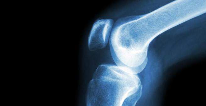
A tibial fracture (tibial fracture) is a fracture of the upper part of the tibial bone. Typical symptoms include pain, swelling and knee joint effusion. The Tibiakopffraktur arises mostly by a jump from high altitude or the action of large forces as in a traffic accident. Learn more about the symptoms, risk factors and treatment here.

Tibialis fracture: description
In a Tibialopffraktur the head of the tibia is broken. Frequently, the knee joint is also involved. Tibial bone fractures account for about one to two percent of all fractures.
Since the leg axis is slightly O-leg-shaped and the outer bone has a thinner bone structure, fractures on the outer side of the tibial bone are more common. Physicians refer to this fracture as a lateral Tibiakopffraktur. Less common is the medial tibial fracture (tibial fracture towards the middle of the body).
In tibial bone fracture, a distinction is made according to the AO classification (AO = Arbeitsgemeinschaft für Osteosynthesefragen) in three different forms:
- A fractures: fractures in which the joint is not affected (bony ligament tear)
- B-fractures: Fractures with partial joint involvement such as cleft fractures, indentation fractures (impression fractures) and impression fractures
- C fractures: complete joint fractures
Tibialis fracture: symptoms
The typical symptoms of a tibial fracture are pain and swelling in the knee and lower leg area. Almost always a knee joint effusion also occurs. This accumulates blood within the joint capsule. In technical terms, this is also referred to as hemarthrosis. Due to the pain, the affected person can no longer properly move the knee joint.
Frequently, the tibialis fracture additionally injures the lateral and transverse ligaments. The meniscus can also be affected. If several bone fragments have arisen or if a fragmentary fracture is present, there is always the danger of a compartment syndrome of the lower leg. The tissue pressure is increased by swelling and blood, which squeezes nerves, muscles and vessels within a fascia. If the tissue is permanently damaged, claw toe can occur.
Tibialis fracture: causes and risk factors
A bony tibia fracture, for example, occurs when the bones are heavily compressed from above – for example, when landing on a high leg on a stretched leg. The bone is compressed so that the head of the tibia splits. Usually both legs are affected. Often, even high-trauma traumas such as traffic accidents (cars, motorbikes) or sports accidents (skiing, cycling) are the cause of a broken tibial bone.
In younger patients, a cleft fracture often occurs, which may be combined with an indentation fracture (impression fracture). In elderly patients, osteoporotic bone is the cause of tibial fracture in many cases. Then usually Eindrückungsfrakturen arise.
Belt injuries in this area are caused by torsional and shear loads. In about 63 percent of cases additionally meniscal and cruciate ligament injuries occur.
Tibialis fracture: examinations and diagnosis
The responsible specialist for a Tibiakopffraktur is a doctor for orthopedics and accident surgery. He will ask you first about the accident and your medical history. Possible questions are:
- How did the accident happen?
- Do you have pain?
- Can you still move the leg or bend the knee?
- Did you already have complaints like pain and restricted mobility?
After that, the doctor will examine your leg closely and watch for soft tissue or other accompanying injuries. Evidence of soft tissue damage is provided by contusion marks, blisters and superficial and deep wounds.
Tibialis fracture: Imaging examination
To further diagnose a Tibiakopffraktur an X-ray is made. The leg is x-rayed from the side and from the front. To plan an operation, computerized tomography (CT) is usually performed. For difficult knee injuries, magnetic resonance imaging (MRI) can be helpful in assessing meniscal and ligament injuries more accurately.
Tibialis fracture: treatment
A tibial bone fracture is first immobilized in a plaster splint or Velcro closure rail to relieve the leg and allow it to swell. In the further course, such a break is rarely treated conservatively. In most cases, surgery is required.
Tibialis fracture: Conservative treatment
Unshifted or minimally displaced cleft and indentation fractures (impression fractures) can be treated conservatively. However, the muscles attaching near the knee joint often pull the bone fragments apart, which later shifts the fracture.
After the first phase has been overcome, the knee joint is usually moved passively with a motor rail. The leg can be weighted from 10 to 15 kilograms with walking sticks and a velcro strap for about six to eight weeks. After another six to eight weeks, the load can be slowly increased to half the body weight.
Tibialis fracture: surgical treatment
All other cases of tibial fracture are usually treated surgically. The aim of the treatment is to restore the articular surface and to begin exercises as early as possible. Simple cracks are bolted. The injured articular surface is filled up – either with the body’s own bone material (from the iliac crest) or synthetically produced bone substitute material such as calcium phosphate or hydroxyapatite.
In heavy debris fractures, visible malposition or an unstable leg, the tibial bone is reoriented under anesthesia and stabilized externally with a fixator. This is a holding system for fixing bone fragments.
After the operation, the knee joint is regularly moved passively with a motor rail. The leg should then be relieved for about six to twelve weeks.
Tibialis fracture: disease course and prognosis
The healing process in a Tibiakopffraktur is different. He is monitored by regular X-ray checks by the doctor. With conservative treatment, it takes an average of eight to ten weeks for the bone to heal. In case of a minor tibial fracture, the long-term prognosis is usually very good. If a tibial fracture is operated, the prognosis also depends on the age of the patient and existing pre-existing conditions such as joint wear (osteoarthritis) and bone loss (osteoporosis).
Tibialis fracture: complications
If the ligaments are involved in the tibial fracture, or if it is a debris fracture, there is always a risk that the artery of the popliteal fossa (A. poplitea) was also injured. The nerves are rarely involved.
After a debris fracture or a difficult indentation fracture (impression fracture), osteoarthritis of the knee (gonarthrosis) can occur.
Other possible complications include wound healing disorders. These often occur in too early surgery, since the tibia is surrounded only by a thin soft tissue shell. Furthermore, an infection can occur: Then the knee joint must be cleared out and thoroughly rinsed. An infection can also be the cause if the tibial plateau fracture does not heal (pseudarthrosis).