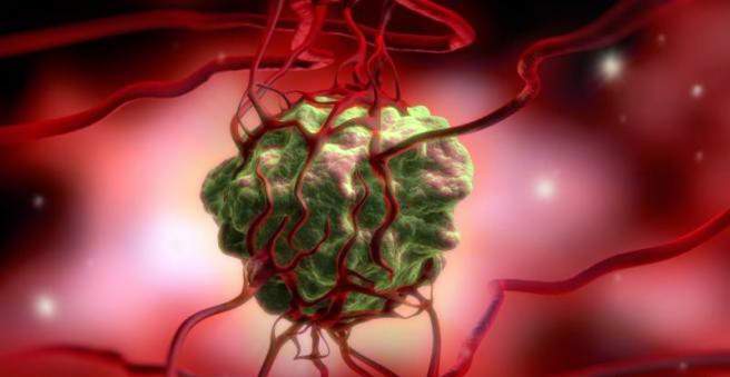
The mesothelioma is a tumor that emanates, for example, from cells of the lung, peritoneal or pericardium and encloses the lungs in the form of a coat. It particularly affects people who have had contact with asbestos for a long time. Mesothelioma can be benign or malignant (pleural cancer, breast cancer). Treatment usually consists of surgery combined with chemotherapy. Learn more about mesothelioma here.

Mesothelioma: description
Mesothelioma is an overgrowth (tumor) of the mesothelium. This is a single-layered epithelial tissue that forms the boundary of body cavities such as pleura, pericardium and peritoneum. It usually lays as a planar tumor around the lungs.
Like most tumors, mesothelioma can appear in benign and malignant forms. The latter is often the result of asbestos exposure. The latency is long: about 35 years elapse between exposure to asbestos and the onset of the first symptoms. If you have been professionally exposed to asbestos and suffer from a malignant mesothelioma, this is considered a recognized occupational disease.
Malignant mesotheliomas are more than 80 percent pleural mesothelioma, ie tumors that emanate from the pleura (pleura: pleura and lung fur). This is referred to as breast cancer or pleural cancer.
About 20 people per million inhabitants in Germany suffer from mesothelioma every year. In many industrialized countries, asbestos was banned, yet the incidence seems to be increasing. Men become three to five times more likely to have mesothelioma than women. The higher the age, the higher the disease risk.
Mesothelioma: symptoms
The pleural cancer symptoms can be very different. It can take up to half a year between the first symptoms and the final diagnosis.
Most sufferers of mesothelioma of the pleura report shortness of breath as the first symptom. In addition, pain in the area of the chest can occur when the intermediate ribs nerves are irritated or the chest wall is infiltrated.
In rare cases, a high diaphragm or a cough may occur. Also rare in pleuropod cancer so-called paraneoplastic symptoms such as anemia, weight loss, fever or a spontaneous pneumothorax (sudden ingress of air into the slit-shaped space between the lung and pleura).
Unilateral pleural effusions or pulmonary thickening with concomitant chest pain may be further evidence of mesothelioma.
Mesothelioma: causes and risk factors
Up to 90 percent of pleural mesothelioma cases can be attributed to asbestos exposure. There has been an asbestos ban in Germany since 1993. Asbestos has been banned in the EU since 2005. Nevertheless, asbestos continues to be massively used industrially worldwide, for example as an insulating material in the construction industry. So far, no limit has been established under which there is no risk of mesothelioma.
About ten to twenty percent of mesothelioma disorders are not caused by asbestos, but for example by zeolite (erionite), an asbestos-like fiber. In addition, other factors are suspected of being able to trigger a mesothelioma. These include, for example, the so-called SV-40 viruses, repeated inflammations and a genetic predisposition to mesothelioma. In addition, experts are currently investigating whether nanomaterials such as nanotubes can also lead to malignant mesothelioma.
Mesothelioma: examinations and diagnosis
If you have signs of pleural mesothelioma, you should consult a GP or pulmonologist. To diagnose a mesothelioma, the doctor will ask exactly the symptoms and your medical history. Typical questions of the doctor would be for example:
- Since when and how often do you have complaints, such as coughing?
- Are you getting bad air?
- Do you have tough expectoration when coughing?
- Has fever also occurred? Do you sweat hard at night?
- Do you have contact with asbestos professionally or privately?
- Do you live or work in the vicinity of astbest processing factories?
- Have you been to areas with natural asbestos deposits?
- Do you live in an old building with asbestos-containing components?
If a mesothelioma is suspected, referral to an experienced lung center is useful. To confirm the suspected diagnosis, further physical examinations follow. To record the size of the tumor, imaging techniques such as ultrasound, computed tomography (CT) and magnetic resonance imaging (MRI) can be performed. Final certainty in suspected mesothelioma brings a histological examination of the altered tissue.
Imaging procedures
To determine whether water has accumulated between the lung and pleura (pleural effusion), the chest is examined by ultrasound (transthoracic ultrasound). A pleural function (see below) is also performed under ultrasound control.
Computerized tomography (CT) is the best way to detect and detect mesothelioma. In addition, it can be determined by means of CT whether the tumor has already formed secondary tumors (metastases) in the lymph nodes.
If it is suspected that the tumor has spread to the diaphragm or chest wall, magnetic resonance imaging (MRI) can be performed. Also, a so-called positron emission tomography (PET) is useful, especially to detect distant metastases.
thoracentesis
In a pleural function, the doctor inserts a fine needle past the ribs into the pleural space and withdraws fluid. In more than half of all patients with pleural cancer, cancer cells can be detected in the pleural effusion. However, a negative result does not rule out a pleural mesothelioma.
needle biopsy
In percutaneous needle biopsy, a needle is advanced into the body from outside to extract a tissue sample from the affected area. The whole is monitored by X-rays, ultrasound, CT or MRI to control the exact position of the needle.
Thoracoscopy (breast mirroring)
To ensure the diagnosis, a thoracoscopy (breast mirroring) is often necessary. The pleural cavity is examined endoscopically. In addition, during the examination, some tumor tissue can be taken for histologic diagnosis.
Fine-tissue diagnostics
The examination of the histological sample should be done by a specialized lung pathologist. The mesothelioma is histologically divided into different forms:
- Epithelial mesothelioma (50 percent of all mesothelioma cases)
- Sarcomatous mesothelioma (25 percent)
- Biphasic mesothelioma (24 percent)
- Undifferentiated mesothelioma (1 percent)
Mesothelioma: treatment
Mesothelioma should be treated in a specialized center because both diagnosis and treatment are challenging. A standardized guideline on how to treat mesothelioma does not exist. However, it is generally known that monotherapy (ie a single method of therapy such as surgery) is not sufficient to combat the aggressive tumor.
Various methods are currently available for the treatment of mesothelioma: Surgical therapy, chemotherapy, radiotherapy and pleurodesis (pectoralis and pleura are surgically combined).
Usually a combination of surgery and chemotherapy and / or radiation is considered to be the most appropriate course of action.
Surgical therapy
Since the pleural mesothelioma often develops multifocal, that is, develops and expands diffusely in several places, only large-scale surgical interventions are useful. Two operative methods are distinguished: pleurectomy / decortication (PD) and extrapleural pneumonectomy (EPP).
Pleurectomy / decortication
During pleurectomy / decortication, only the lung lining, ie the pleura, is removed. The lung itself is preserved. Depending on the size of the tumor, the pericardium and the diaphragm are also removed in some cases.
The advantage of this less radical method is that the patient recovers faster. Since this method does not remove the entire cancerous tissue and tumor tissue remains in the body, there is a high probability that a new mesothelioma will form (recurrence).
Extrapleural pneumonectomy
In young patients with a good general condition, a so-called extrapleural pleuropneumonectomy may be useful. It is the more radical method of removing the lungs along with the lung and pleura, and the diaphragm on the affected side. The diaphragm is reconstructed using Gore-Tex-like material.
Extrapleural pleuropneumonectomy is a major five to eight-hour operation. It severely limits the patient’s performance. The operation should therefore be performed only in early stages of mesothelioma and only at specialized centers.
chemotherapy
In chemotherapy, the mesothelioma is treated with the help of cytostatics, which are administered via the vein at regular intervals. Induction chemotherapy is distinguished from adjuvant chemotherapy. In induction chemotherapy, a high dose of cytotoxic drugs is given at the beginning of treatment. In about one third of those affected, this causes the mesothelioma to partially recede. Adjuvant chemotherapy is performed following surgical therapy. It shows similar success rates.
For chemotherapy, a combination of the two cytostatic agents cisplatin and pemetrexide is usually used. Thus, the best chances of survival and the best quality of life could be achieved.
radiotherapy
Irradiation (radiation therapy, radiotherapy) is used as a preventive measure in patients with mesothelioma in the area of puncture channels and after surgery so that there is no local recurrence. In addition, radiation can help to reduce the pain. In general, however, is from a radiation dispensed with, since the tumor usually spreads complex and thus requires a high dose of radiation. The risk of additionally damaging the lungs and heart is too high.
pleurodesis
In bad general condition and advanced disease, talc pleurosis is useful. The pleura (lung and pleura) are glued together with talcum powder. If there is fluid in the pleural space (pleural effusion), the symptoms can be significantly improved. A pleurodesis can also be performed as part of a diagnostic breast mirroring (thoracoscopy).
Mesothelioma: disease course and prognosis
Today, mesothelioma, despite all available means, can only be cured in exceptional cases. Several factors influence prognosis on a case-by-case basis, including age, gender, tumor subtype, and tumor stage. For example, epithelial mesothelioma has a more favorable prognosis. Likewise, a more favorable prognosis have younger sufferers (under 75 years) and women.
For example, a role in the prognosis also plays a role in whether the patient is restricted in his activity, can still take care of himself and determine himself (Karnofsky Index). Factors such as low hemoglobin levels, high levels of LDH (“bad” cholesterol) or high levels of white blood cells (leucocytes) and platelets (platelets) can also affect prognosis.
Pleural mesothelioma: life expectancy
Survival time in pleurisy is generally four to twelve months. Only twelve percent of those with negative prognosis factors (such as old age) survive the first year.
aftercare
After completing the therapy should mesotheliomaPatients go to the medical control about every two to three months. Attention is paid to tumor-associated complaints and a physical examination is carried out.