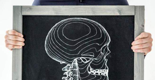
A skull base fracture (temporal bone fracture, otobasal fracture) is a fracture of the skull base due to trauma. Typical symptoms include bruising around the eyes, facial nerve palsy and hearing impairment. The cause of a skull base fracture is a direct trauma to the skull. The treatment is mostly conservative. Find out more about the skull base break here.

Skull base fracture: description
A skull base fracture is commonly seen as a dangerous injury, but usually not because of the fracture itself, but because of the often simultaneously injured brain.
The skull base fracture counts as well as the Kalottenfraktur (break of the skull roof) and the facial skull fracture to the skull fractures.
The most important fracture forms in skull base fracture are:
- Partial temporal bone tear (nose and skull base)
- Temporal bone fracture (ear and skull base)
Occipital fractures occur with 70 to 80 percent of cases significantly more often than the temporal bone fracture. The rupture runs along the longitudinal axis of the temporal bone (os temporale), through the roof of the middle ear space and the facial nerve canal (facial canal). The ossicles often move, which disturbs the sound conduction. There is also the danger that an infection can arise via the external auditory canal. This is the case in about 20 percent of all temporal bone fractures.
In the temporal bone fracture fracture begins at the posterior surface of the petrous pyramid, crosses the roof of the inner ear canal and also moves in the direction of facial nerve canal and / or labyrinth.
Skull base fracture: symptoms
In skull base fracture the symptoms are different, depending on whether it is a longitudinal or transverse fracture of the petrous bone. Since numerous nerves and vessels can pass through the skull base and be injured by the fracture, different failures occur.
Temporal bone fracture (frontobasal fracture)
Bruising around the eyes (monocular or glandular hematoma) is typical of this form of skull base fracture. The eyelids swell up strongly. If the floor of the eye socket is broken, the eyeball can kick deeper (blow-out fracture), whereby the patient usually sees double images.
When the temporal bone fracture also the paranasal sinuses are injured. It can also form steps in the external auditory canal. In addition, the eardrum can rupture, the ossicular chain can be interrupted and the sound conductor may be disturbed.
In 15 to 25 percent of cases of temporal bone fracture, the facial nerve is paralyzed. By tearing off the olfactory nerves, the sense of smell is disturbed. Nervous water or blood can drain from the nose, ear or mouth.
Partial transverse fracture (laterobasal fracture)
The lateral fracture usually affects the ear: Due to the damage to the inner ear of the affected person can hear nothing more and the sense of balance falls out. Facial nerve paralysis occurs much more frequently than in heel bone fracture. About half of the patients with temporal bone fracture are affected. Nerve water can also escape via the ear. This is why a bruise behind the ear, also called “battle’s sign”, is often found in the lateral fracture of the petrous apex.
Skull base fracture: causes and risk factors
The skull base fracture is caused by a strong impact on the skull, such as in the context of traffic accidents or brawls. More than half of the victims with skull base fracture had a traffic accident, mostly with a frontal impact.
In about 17 percent of all patients with fracture of the skull roof, the fracture gap reaches into the skull base. Skull fracture generally occurs together with craniocerebral trauma (SHT). In about four percent of all patients with severe craniocerebral trauma, there is an isolated skull base fracture. Due to the swelling in the facial area and because other consequences of craniocerebral trauma are usually in the foreground, the skull base fracture is often not noticed.
Skull base fracture: examinations and diagnosis
Affected with skull base fracture are often injured multiple times (polytrauma) and are initially in the intensive care unit. In order to diagnose a skull base fracture, the doctor will first ask the patient – as far as his condition allows – about the accident and his medical history. Some questions from the doctor might be:
- How did the accident end?
- Do you have pain?
- Did you notice that fluid leaked from your ears, mouth or nose?
- Do you have problems speaking, hearing or seeing?
This is followed by physical examinations. In a complex skull base fracture usually work together several doctors from different departments such as neurosurgery, oral and maxillofacial surgery and ENT surgery together.
The doctor examines the external auditory canal of the patient, paying attention to whether a step or ear secretion has formed. If the eardrum is still preserved, usually blood accumulates in the middle ear (hematotympanum). Then, if possible, the auditory function is checked.
A middle ear deafness can already be distinguished with the tuning fork of an inner ear hearing loss. The balance is judged with the so-called Frenzel glasses. If the organ of balance fails, it causes nystagmus.
Next, the doctor will examine the basal cranial nerves and the large vessels. In facial paralysis, it is important to distinguish whether the paralysis has developed gradually or has been fully developed from the start. This helps to estimate the prognosis and to plan the further course of action.
If the affected person loses cerebrospinal fluid (CSF) or blood from the nose, ear or mouth, this may also be an indication of a skull base fracture. Since leaking nerve water from the nose looks very similar to nasal secretions, a laboratory examination is necessary. Special test strips are used, which determine the sugar concentration (glucose concentration): In the nervous water, the sugar concentration is higher than in the nasal secretions.
Skull base fracture: Apparative diagnostics
The above mentioned hints can arouse suspicion of a skull base fracture, but do not prove it. Even in the conventional X-ray, a skull base fracture is difficult to detect. Further diagnostics are therefore always carried out with computed tomography (CT). If the physician discovers air in the interior of the skull or air-filled bone compartments such as paranasal sinuses and mastoidal cells (pneumoencephalon) on the images of the brain and facial skull, this indicates a skull base fracture. A fracture gap does not necessarily have to be visible.
If the affected person has lost their hearing or facial paralysis, magnetic resonance imaging (MRI) is used to exclude any possible bruising in the brain and visualize the facial nerves.
Skull base fracture: treatment
Patients with skull base fracture must be monitored during the first 24 hours. In addition, the treatment depends on the extent of the skull base fracture. In most cases, conservative treatment takes place. However, if bone components are shattered and displaced or there are passages where nerve water escapes, surgery becomes inevitable. Even with an immediate, total facial paralysis (facial palsy) must be operated on. The procedure is usually performed only after the swelling in the brain has decreased again. Exception is a space-demanding bruise.
The injured ear canal is cleaned and covered under sterile conditions. If the skull base fracture has led to an inner ear deafness, a so-called rheological treatment is initiated as in the case of an acute hearing loss: Certain substances are used to improve the perfusion in the inner ear. Occurring dizziness can be alleviated with so-called Antivertiginosa.
If nerve water escapes from the nose, ear or mouth as a result of the skull base fracture, it must first be treated with antibiotics as a preventive measure to avoid an ascending infection. If the defect is located in the middle fossa, with nerve water draining through the ear, this gap usually spontaneously adheres and seldom needs surgical treatment.
Nerves that are trapped in the fracture line do not necessarily require surgery. Sometimes they can regenerate spontaneously.
Skull base fracture: surgery
In fractures in the anterior fossa, especially the cribriform plate, when nerve water drains away from the nose, surgery is always required. Because the gap does not close spontaneously, and even years later, an infection can develop. During the operation, the meninges (dura) are first closed in a water-tight manner. Thereafter, the bone is reconstructed.
Bleeding caused by torn cerebral vessels must also be stopped surgically. Here, the bruise, which is located in the so-called epidural space, must be cleared. This prevents the pressure in the brain from increasing and causing brain damage.
Skull base fracture: disease course and prognosis
In skull base fracture, the prognosis differs depending on the type of fracture. A longitudinal fracture usually has a good prognosis and rarely causes consequential damage. A transverse fracture in which the inner ear function and the facial nerve have been damaged, however, is expected to be permanent damage.
Skull base fracture: complications
Possible complications of a skull base fracture are:
- Brain inflammation (meningitis)
- Pus accumulations (empyema)
- Brain Abscess
- Carotid artery injury (Arteria carotis)
- Carotid-cavernous sinus fistula (vessel short-circuit through which blood flows from the carotid artery into the venous network in the skull)
- permanent cranial nerve lesions
Such complications can increase the prognosis at one Fracture of the base of the scull deteriorate.