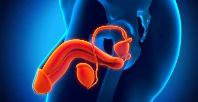
The epididymitis (epididymitis) is a painful inflammation of the epididymis. It must be treated. A delayed inflammation can lead to infertility. The mostly multi-week treatment of epididymitis includes bed rest, painkillers and possibly also antibiotics. Read all important information about symptoms, diagnostics and treatment of epididymitis!

Epididymitis: description
The epididymitis (epididymitis) arises in most cases as a concomitant of bacterial inflammation of the prostate or urinary tract. It rarely occurs in isolation as the sole inflammation of the epididymis, as the pathogens spread along the vas deferens. The epididymitis usually occurs only from puberty on.
The epididymitis can be acute or chronic.
Testes and epididymis
The epididymis counts – as well as the penis and the testicles – to the outer sexual organs of the man. The testicles and epididymis lie together in the scrotum (scrotum). The testicles produce the semen and the sex hormones. At the upper pole of the testicle, the fresh seed is passed into the epididymis and stored there. The vas deferens emerges from the epididymis and then flows into the area of the prostate gland into the urethra.
Although testicles and epididymides are close together and are closely related, orchitis is not the same as epididymitis. The latter is much more common. In some cases, however, the inflammation affects both testes and epididymides. In this case we speak of epididymorchitis.
Epididymitis: symptoms
The symptoms of an epididymitis similar to those of an orchitis (Orchitis): Usually it comes relatively suddenly to a painful, partially palpable swelling of the scrotum, what doctors call “acute scrotum”. The surrounding skin shows typical signs of inflammation such as overheating and redness. The epididymal pain can radiate into the groin and lower abdomen. The accompanying symptoms include fever up to 40 ° C as well as vomiting and nausea.
The chronic epididymitis may also be characterized by a painless swelling. The epididymitis caused by chlamydia can be relatively symptom-poor.
Epididymal inflammation: causes and risk factors
The most common causes of epididymitis include bacterial inflammation of the urinary tract and the prostate. As a rule, the pathogens from the urethra or the prostate reach the epididymis via the vas deferens. One then speaks of an “ascending (ascending) infection”.
Men with bladder bladder dysfunction, urogenital malformations and a permanent urinary bladder therefore have a particularly high disease risk. In children, malformations of the urinary tract are responsible for the bacteria getting into the epididymis. In some cases, a testicular torsion, that is a twisting of the testicles, leads to an epididymal inflammation. Epididymis are in many cases but not isolated inflamed, but together with the adjacent sections of the seminal and urinary tract.
Which pathogens trigger the inflammation?
A testicular inflammation is usually triggered by viruses – not so the epididymitis. Cause here are usually bacteria. This is often the case of Chlamydia trachomatis (rare Neisseria gonorrhea) among men under 35 (sexually active). Intestinal bacteria such as Escherichia coli, Enterococci, Klebsiella or Pseudomonas aeruginosa as well as staphylococci are responsible for epididymitis in men over the age of 35 years.
Epididymitis is rarely caused by a spread of bacteria via the bloodstream (especially pneumococci and meningococci), as part of a tuberculosis disease or through trauma: If urine flows into the seminal veins, chemical irritation of the epididymis may occur causes an inflammatory process.
Other possible causes
The rarer viral inflammation of the epididymis is usually from the mumps virus. In this case, the testes are often affected, with the epididymitis may precede the orchitis. Before adolescence, adeno- and enteroviruses can trigger a so-called post-infection, as a post-infection epididymitis.
Autoimmune processes are also considered as a cause of epididymitis.
Fungi (Candida, Coccoidioides, Histoplasma etc.) and worms (Schistosoma, Wucherichia or Echinococcus) are rare causes of epididymitis in Germany.
In addition, there is an isolated description of epididymitis, which were triggered by drugs such as amiodarone (remedy for arrhythmia).
Epididymitis: examinations and diagnosis
If you suspect an epididymitis you should contact a urologist. The doctor will first talk in detail with you about your complaints and possible underlying diseases (medical history.) Possible questions are for example:
- Since when do the complaints exist?
- Did the symptoms start suddenly?
- Do you have discharge from the penis or pain when urinating?
- Are you already aware of diseases of the urinary tract (including urinary tract infections)?
- Do you have intercourse?
Epididymitis: physical examination
Following is the physical examination. The doctor will first examine the scrotum for recognizable signs of inflammation (overheating, redness) and check whether the epididymis is swollen.
Then the doctor raises the scrotum. If this reduces the symptoms (Prehn sign positive), this speaks for epididymitis. With a testicle inflammation and a testicular torsion (rotation of the testicle around its own axis) the complaints on the other hand by the lifting of the scrotum do not get better. This distinction is very important because testicular torsion is an emergency that needs to be operated on within a few hours. In testicular torsion, however, an inflammation of the epididymis may also occur as a concomitant. If testicular torsion can not be excluded during the examination, an operative exposure of the testicle is necessary. If an abscess (encapsulated collection of pus) has already formed in the area of the epididymis, it can be palpated as a fluctuating swelling.
Epididymal inflammation: laboratory tests
The doctor will also ask you for a urine sample. On the one hand you can quickly confirm the suspicion of an urinary tract infection with so-called “urine stix” and on the other hand create so-called urine cultures. The latter should help to determine the causative agent and its sensitivity to certain antibiotics (Resistogramm). In addition, if epididymitis is suspected, a smear of the urethra entrance can also be taken and examined in the laboratory.
In the case of an epididymitis, blood tests show typical signs of inflammation (such as an increased number of white blood cells). Suspected mumps virus infection can be used to detect antibodies in the blood.
Epididymal disease: Imaging
If an epididymitis is suspected, the ultrasound examination of the testicle (testicular ultrasonography) is particularly important for diagnosis. It can be repeated at any time and is completely safe. Therefore, sonography is also excellent for assessing the course of the disease. The urologist recognizes on the ultrasound image the extent of the inflammation and whether the process has already spread to the adjacent testicles. Even a beginning abscess formation can be recognized in time.
If there is a suspicion of obstruction in the urinary system, which urges the urine into the vas deferens and testicles, ultrasound examinations and possibly also x-ray examinations of the urinary tract are performed with a contrast agent (urography). For example, bottlenecks in the urethra (urethral strictures) can be identified. Optionally, a measurement of the urinary jet or a bubble mirror may be necessary.
Epididymal inflammation: treatment
Therapy of an epididymitis consists of bed rest, analgesics and possibly antibiotics. It is important to store the testicles and to cool them with cold compresses. The acute inflammation can last for eight to ten days. The healing process is characterized by the normalization of the temperature, the disappearance of the pain and the slowing down of the epididymis. Only then can the patient get up. He receives a jockstrap (a bag-shaped bandage to protect the testicles), so that epididymis and testicles can not fall.
In severe pain, the spermatic cord can be infiltrated with local anesthetics. During bed rest, there is an increased risk of thrombosis. For the prevention of blood clots, the patient may therefore be given anticoagulant heparin.
In children, a malformation of the urinary tract, which obstructs the urinary outflow, usually leads to an epididymitis. In order to get the healing going faster, the urine is often temporarily removed from the bladder (puncture cystostomy). If necessary, surgical treatment of the malformation is necessary following the treatment of epididymitis.
If an abscess (encapsulated collection of pus) forms as a result of epididymitis, it must be surgically opened and removed.
If an infection with chlamydia triggers the epididymitis, all sexual partners should always be treated. Otherwise, repeated infections (re-infections) are possible.
In the case of a chronic course of treatment takes longer (especially the antibiotic dose). In severe cases, the epididymis must be surgically removed (epididymectomy) or the spermatic cord severed (vasectomy).
If the spermatozoa secrete due to inflammation (occlusive azoospermia), this can be remedied with the help of microsurgical techniques after the inflammation has subsided. Within the so-called epididymovasostomy, a continuous pathway for the spermatozoa is created again.
Epididymal disease: disease course and prognosis
A lot of patience requires the therapy of an epididymitis: duration of the healing process can be up to six weeks – even with optimal treatment. Only then does the scrotum feel normal again in many men.
As a rule, epididymitis heals well. But there are also complications possible such as fistulas, local destruction of the epididymal tissue and a transmission of inflammation along the seminal and urinary tract. Occasionally, the inflammatory focus (abscess) encapsulates in a pronounced epididymitis. He then has to be eliminated surgically.
Frequent or delayed epididymal inflammation can lead to scarring and constrictions in the epididymis or vas deferens. As a result, the transport of sperm can be hindered, which leads to infertility, especially in a bilateral closure (occlusive azoospermia). In addition, the inflammation may spread to, among other things, the adjacent testes.
For recurrent epididymitis, often only an operative separation of the spermatic cord (vasectomy) or the removal of the epididymis (epididymectomy) helps. In addition, in the advanced stages of inflammation sometimes the testicles have to be removed.
In addition to blood poisoning (sepsis), the so-called Fournier gangrene is a dreaded complication when the Epididymitis in a weakened immune system is very difficult. This leads to a tissue dehydration (necrosis) of connective tissue strands in the testes, which can lead to a severe inflammatory reaction of the entire organism with a high mortality.