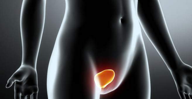
Bladder stones are urinary stones in the urinary bladder. They usually form in the urinary bladder itself, for example, when the urine can not flow freely when urinating. In addition, urinary stones from the renal pelvis can be transported via the ureter into the bladder. In many cases, bladder stones are flushed out of the body on their own, but sometimes they have to be removed surgically or with special techniques. Read all important information about bladder stones here.

Bladder Stones: Description
In general, a urinary stone is a solid, stone-like concretion in the urinary tract. If there is a urinary stone in the urinary bladder, this concretion is called a bladder stone. The urinary bladder collects the urine as a reservoir and allows it to be given voluntarily by special musculature. Bladder stones can either form themselves in the bladder (primary bladder stones) or they form in the kidney or the ureters and finally enter the urinary bladder (secondary bladder stones) with the constant flow of urine. The urinary stones symptoms are the same in both species.
A blister stone is formed when certain rock-forming salts crystallize out in the urine. This usually happens when the salt in question is in too high a concentration in the urine and thus exceeds the solubility threshold. If the salt forms a solid crystal (concretion), over time more and more layers deposit on it, so that the initially small calculus becomes an ever-growing urinary stone.
Depending on the type of salt that makes up the stone, physicians distinguish:
- Calcium oxalate stones (75 percent of all urinary stones)
- “Struvite Stones” made of Magnesium Ammonium Phosphate (10 percent)
- Urate stones from uric acid (5 percent)
- Calcium Phosphate Stones (5 percent)
- Cystin stones (rare)
- Xanthin Stones (rare)
The distinction of the different stone types is not only for purely scientific reasons. Rather, the different stone types differ in terms of their causes, diagnostics and treatment. Thus, for example, only the calcium-rich, “radiopaque” stones detected on the X-ray or only certain urinary stones with an alkalization of urine may be resolved again.
Bladder stones can occur in people of all ages. However, older and overweight people are more prone to bladder stones. Men and women are equally affected. In men, the cause of bladder stones is benign enlargement of the prostate (BPH) in most cases.
In many cases, bladder stones cause no discomfort and are flushed out of the body by itself with the urine. However, if the urinary stones block the exit to the urethra or are too large to pass through the urethra by themselves, medical urinary stone removal is necessary. Here, urinary stones can be crushed during a bladder reflex with a pair of pliers or by means of the so-called shock wave therapy (ESWL). The resulting chunks are then small enough to be flushed out with the urine stream. Correct surgery is only necessary in a few cases with very large bladder stones. In addition to the removal, especially the removal of causes is important in order to prevent re-bloating stones.
Bladder stones: symptoms
People with bladder stones often have no complaints. Whether the bladder stones cause symptoms depends primarily on where the stone is exactly and how tall it is. If it is free in the urinary bladder, the urine can flow unhindered through the urethra (urethra). Special symptoms do not occur in this case. On the other hand, if he sits firmly against the lower bladder wall and obstructs the exit of the urinary bladder through the urethra, discomfort develops. The symptoms arise on the one hand by the mucous membrane irritation, which is caused by the often sharp-edged bladder stone and on the other hand by the urine, which often accumulates up to the kidney. Typical bladder stone symptoms are sudden, colicky pelvic pain that can radiate to the flanks. In addition, when urinating pain, the urinary stream can suddenly stop and the urine bloody. Often there is also a constant urge to urinate, combined with a small amount of urine during urination (pollakisuria).
How strong the symptoms are depends on how big the bladder stone is. Smaller urinary stones usually lay the opening to the urethra only partially and still allow a certain amount of urine. With larger stones, less and less urine can pass through the urethra, so the symptoms usually increase as the size of the stone increases. A complete obstruction of the urethra leads to a build-up of urine in the bladder, which can reach over the ureters to the kidneys. This situation, where urine delivery is no longer possible, is referred to by physicians as urinary retention or ischuria.
Many sufferers show an increasing agitation in addition to these symptoms. This comes mainly from the fact that those affected unconsciously look for a body position in which the pain subsides. Thus, they constantly change from lying to a standing position or go around. In addition, nausea and even vomiting may occur as a result of the pain.
If you notice pain when you urinate or unusual, spasmodic pain in the lower abdomen, it is best to see a doctor immediately and clarify the cause. If the urine builds up to the kidneys, it can permanently damage the kidneys.
Bladder stones: causes and risk factors
Bladder stones are mineral salts that are normally dissolved in the urine and are flushed out of the body with it. These mineral salts may, under certain circumstances, be released from the urine (they “precipitate”) and settle in the bladder. At the beginning of development, bladder stones are very small, crystal-like structures. By the addition of further salts they often grow steadily.
Doctors differentiate primary and secondary bladder stones, Primary bladder stones are produced in the urinary bladder itself, secondary bladder stones are formed in the upper urinary tract organs, such as the kidney or the ureters, and are flushed into the bladder with the urine. However, primary bladder stones are much more common than secondary bladder stones. If the urinary stones from kidney or ureter dissolve, they are usually so small that they are easily eliminated and do not get stuck in the bladder.
Mostly bladder stones are formed when urine discharge from the bladder is obstructed (primary bladder stones). The urine lingers for an excessively long time in the bladder, so that it comes to the precipitation of mineral salts and thus to urinary stones. Frequently, this also causes inflammation of the urinary tract, which in turn favors the formation of bladder stones.
The typical causes of urinary obstruction include prostate enlargement or a neurogenic bladder voiding disorder: Benign enlargement of the prostate (BPH) is a very common finding in older men. Also in neurological diseases such as multiple sclerosis or paraplegia may occur due to drainage disorders to the formation of bladder stones. In these diseases, the contraction of the bladder muscles and thus the urination (micturition) is often impaired.
In a urinary tract infection, the bacteria can change the chemical composition of the urine and increase the risk of precipitation of certain substances. Thus, the formation of magnesium ammonium phosphate struvite stones is attributed to urinary tract infections with certain bacteria.
In Germany, an unfavorable diet with lots of animal fats, proteins and oxalic acid foods is considered a risk factor for the formation of bladder stones. Oxalic acid is included in nuts, coffee, cocoa, rhubarb, beetroot and spinach, for example. Stone-forming substances such as oxalate, calcium, phosphate, ammonium and uric acid (urate) can only be dissolved in the urine in a certain amount and transported out of the body. If the amount taken up with food exceeds a certain limit, this may also lead to the precipitation of certain substances.
Risk factors for bladder stones are also foreign bodies in the bladder, such as bladder catheter or surgical sutures. Bacteria are particularly easy to adhere to foreign bodies and thus trigger a urinary tract infection. The infection in turn increases the risk of bladder stones.
Other risk factors for bladder stones are:
- too low fluid intake (concentrated urine)
- one-sided diet with too much meat and dairy products
- increased intake of vitamin D3 (for example, vitamin capsules)
- Lack of vitamin B6 and vitamin A.
- Osteoporosis with increased calcium release from the bones into the blood
- Parathyroid hyperfunction (hyperparathyroidism) due to increased levels of calcium in the blood associated with this condition
- too high magnesium intake
Bladder stones: examinations and diagnosis
If you suspect bladder stones, a specialist in urinary disorders (urologist) is the right person to contact. In large cities, there are also established urologists with their own practice, in rural areas, urologists are usually only found in hospital. First, the attending physician is the medical history (anamnese) raise. In doing so, you describe to the doctor your current symptoms and any previous illnesses. Afterwards, the doctor will ask you further questions to better understand your personal case. These can be questions like:
- Where exactly are you in pain?
- Do you currently have problems urinating?
- Did you have problems urinating even before the symptoms started?
- Is an enlarged prostate known to you (men)?
- Did you notice blood in the urine?
- Do you take any medicine?
After the anamnesis follows the physical examination, The doctor, for example, listens with the stethoscope on his stomach and then carefully scans it. The physical examination helps the doctor to better assess the causes of abdominal pain and what further examinations are necessary for clarification.
Further investigations:
In case of a suspected bladder stones more examinations are usually necessary. For this purpose, if the patient is able to let water despite the bladder stone, the urine is examined in the laboratory for crystals, blood and bacteria. In addition, a blood sample is taken with which the renal function can be estimated and the uric acid value can be determined. A blood count and blood clotting indicate possible concomitant inflammation in the bladder. In the case of inflammation in the body, the value of the white blood cells (leukocytes) and the so-called C-reactive protein (CRP) in the blood are greatly increased.
An x-ray or ultrasound examination (sonography) can visualize urinary stones. In the radiograph, however, only the so-called “radiopaque” (calcium-containing) stones are clearly visible. Another possibility with which even radiolucent stones can be displayed is urography. A contrast agent is injected into a vein. This is distributed in the body and makes it possible to visualize the kidney and the urinary tract with possible stones. In the meantime, however, urography has largely been replaced by computed tomography (CT). With a computer tomography, all types of stone and a possible urine jam can be detected safely and quickly.
Another method of examination is cystoscopy. In this case, a rod or catheter-like instrument with integrated camera (endoscope) is introduced into the bladder. Thus, stones can be detected directly on the transmitted live images. The advantage of cystoscopy is that smaller stones can be removed beautifully during the examination. In addition, other causes of blockage of urinary outflow from the bladder, such as tumors, may be recognized.
Bladder stones: treatment
If pain persists, the first step in the treatment is the administration of a painkiller. In many cases, a thorough examination is possible only through the previous pain relief. Even symptomless bladder stones, which are discovered by chance during a routine ultrasound, should be treated, as they can increase in size over time and then cause discomfort.
It depends primarily on the size and location of the bladder stone, whether it must be removed or waiting for a spontaneous departure. In most cases, a bladder stone requires no special treatment. Small (≤ 5 mm) and free-lying stones in the urinary bladder are flushed out of the urethra in about 90 percent of the cases. The drainage can be relieved by certain drugs (for example, tamsulosin), for example, when an enlarged prostate narrows the urethra. With some stones (Uratsteinen, Cystinsteinen) one can also try to dissolve the urinary stones by a chemical reaction, or to reduce them (chemolitholysis).
In any case, it is important that you drink a lot to make it easier for the stone to escape. If pain occurs (as is often the case when the urinary tract passes through the urinary tract), analgesics such as diclofenac can help.
If the stone is too large for a spontaneous finish, the stone closes the urethra and thus there is a urine jam, as well as if there is evidence of a serious infection (urosepsis), the stone must be surgically removed. Smaller stones, the doctor in a cystoscopy (cystoscopy) with a pair of pliers or directly remove. In the case of cystoscopy, adults only need local anesthesia, so you can follow the procedure on a monitor yourself. In children, the procedure is performed under general anesthesia. After blistering, you may either return home the same day or within the next two to three days.
How long you need to stay in the hospital after treatment depends on how large the stone was removed and whether complications occurred during the procedure. As with any surgical procedure, there are risks associated with cystoscopy. In general, there is the risk that germs are introduced into the bladder with the instruments and these become inflamed. In addition, organ walls can be injured or even pierced with the instrument. Such incidents are very rare.
For some years now, most of the interventions have involved the crushing of stones by pressure waves. This procedure is called extracorporeal shockwave lithotripsy (ESWL). In the ESWL larger stones are destroyed by shock waves, so that the debris (now much smaller) can be easily eliminated through the urine. If pain persists after removal of the bladder stone, this may be an indication of inflammation of the urinary bladder (cystitis). This is optionally treated with antibiotics.
An open operation method is used today only in very rare cases. It is necessary, for example, if the doctor does not enter the bladder with the endoscope when the bladder is being mirrored because the stone or other structure blocks the urethra or the entrance to the bladder. For example, tumors in the computerized tomography image can sometimes look like urinary stones as well. However, tumors basically require a completely different treatment method, so that in case of doubt, surgery is more open.
If the bladder stones were caused by a disturbance of the bladder emptying, then the treatment of the cause is in the foreground after the stone removal. In men, prostate enlargement often leads to urethral discharge disturbances and subsequent stone formation. In such a case, one can first try to treat the prostate enlargement drug. In the case of a greatly enlarged prostate or recurring urinary stones, however, a surgical intervention is recommended to eliminate the trigger of the formation of stones. A so-called transurethral prostate resection (TURP) is usually recommended. In this procedure, the prostate is removed through the urethra.
Bladder stones: disease course and prognosis
About 90 percent of the bladder stones, which are ≤ 5 millimeters, are flushed out with the urine on their own. Meanwhile, however, severe pain may occur as the bladder stone “wanders” through the urethra. As a rule, all urinary stones that do not go away by themselves can be removed with an interventional or surgical intervention. Basically, you first try to wait for a spontaneous stone finish before considering an intervention.
Consequential damage from bladder stones is rare, for example when a sharp bladder stone injures the bladder wall or the urethra. If the stone wanders through the urethra, it can literally “slit” the wall of the urethra. This can lead to scarring of the urethra and thus permanent urinary problems.
Successful bladder stone removal does not guarantee that there will be no further blemishes after that. Doctors repeatedly point out that urinary stones have a high recurrence rate. This means that people who once had bladder stones are at risk of developing them again.
You can reduce the risk of bladder stones by paying attention to regular exercise and a balanced diet that is high in fiber and low in animal protein. Especially if you have ever had bladder stones, you should only take purine and oxalic acid foods in small quantities. These foods include meat (especially offal), fish and seafood, legumes (beans, lentils, peas), black tea and coffee, rhubarb, spinach and Swiss chard. In addition, you should be careful to drink at least 2.5 liters per day, as well as the urinary tract flushed through, and thus the risk that mineral salts can settle, drops. A safe method bladder stones However, there is no such thing as a general avoidance.