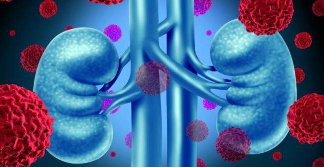
A renal infarction is understood to mean the destruction of kidney tissue because it is no longer adequately perfused by occlusion of the renal vessels. A typical symptom is the acute colic-like flank pain. If the kidney infarction is noticed in time, it is easy to treat and severe consequences are rare. Here you read all important information about renal infarction.

Kidney infarction: description
Similar to a heart attack, kidney infarction occurs when a kidney’s blood vessel closes, usually through a blood clot, and the corresponding kidney tissue is no longer adequately supplied with oxygen. If the blood circulation is not restored in a very short time, it is destroyed. Thanks to good precautionary measures, renal infarction is a rare event. In a few cases renal infarction leads to acute renal failure.
The kidney has in our body the crucial task to cleanse the blood of salts and waste. The renal arteries descend from the abdominal aorta at the level of the second lumbar vertebra and branch into two or three strains. These form so-called end vessels, which means that there are no short-circuit connections (collaterals) between adjacent blood vessels.
Complete renal infarction and partial kidney infarction
Depending on the extent one differentiates a complete renal infarction from the renal partial infarction. In complete renal infarction, the endartery is completely closed. In partial kidney infarction, the renal vessel is either partially occluded, or adjacent bloodstreams have formed over a slow narrowing. This can happen, for example, if a thrombosis, ie a blood clot in the renal veins, has developed slowly. Parallel blood vessels can then prevent such infarction.
In a complete renal infarction, the affected kidney tissue is destroyed within one to two hours (necrosis). If the renal vessel is only partially occluded, or if there are adjacent bloodstreams (collateral blood flow), the kidney can still be rescued if blood circulation can be restored within 24 to 48 hours.
Ischemic and hemorrhagic renal infarction
Kidney infarction can be caused by occlusion of a renal artery or vein.
If a renal artery is affected, it is a so-called ischemic renal infarction. Depending on where the closure is, different forms are distinguished. In wedge-shaped renal infarction, the smallest arteries (arteriae interlobulares) are affected by the occlusion; in trapezoidal renal infarction, the next largest arteries (arteria arcuatae). If the constriction is in the renal artery strain, renal infarction usually spreads in half or all of the kidney.
In the case of hemorrhagic infarction of the kidney, a renal vein is affected by the occlusion. Because the blood can no longer flow away, a congestion develops and fresh oxygen-rich blood can no longer flow.
Renal infarction: symptoms
Renal infarction may manifest itself in different ways. The cardinal symptom is violent, sudden and non-colic flank pain. In case of a major renal infarction, the person complains additionally about abdominal pain as well as nausea or vomiting. In the following days, blood in the urine can be seen as a sign of acute renal failure (gross hematuria).
However, a small renal infarction may also go unnoticed and is only noticeable through poor kidney function. About 25 percent of all cases of renal infarction are due to incomplete occlusion without any symptoms. In most cases, surgery or angiography of the renal vessels is the cause of renal infarction. It is therefore an embolism with arteriosclerotic material, which can be deposited not only in vessels of the kidney, but also in other organs. Depending on which organs it affects, additional symptoms and events such as visual field defects, muscle pain, acute inflammation of the pancreas (pancreatitis) or splenic infarctions may occur.
With a cholesterol embolism so-called gangrenous changes in the area of the toes can occur: tissue dies off and darkens due to the slow degradation of the red blood pigment.
Renal infarction: causes and risk factors
The main causes of renal infarction are embolism, but thrombosis is also an option. In both cases, a renal vessel is blocked by a blood clot: In embolisms, this blood clot from another body region (usually from the heart) was washed ashore. In thrombosis, the clot (thrombus) develops on site.
Kidney infarction due to an embolism
Kidney infarction is most commonly caused by embolism. The blood clot (embolus) usually comes from the heart, eventually gets stuck in a small renal artery and clogs it up. The embolus comes in detail:
- from the left atrium of the heart: in atrial fibrillation
- from the left ventricle: These are blood clots of the heart wall and deposits (vegetations) in bacterial endocarditis (inflammation of the heart’s lining).
- from the main artery (aorta): The so-called arteriosclerotic plaques (inflammatory changes in the blood vessels) may become detached during procedures on the aorta (such as a cardiac catheterization) or vascular surgery. They usually clog both renal vessels.
About 20 percent of the cardiac output, which is the volume of blood pumped from the heart into the bloodstream in a minute, flows through the kidney. Therefore, it is understandable that blood clots can easily enter the renal vessels and trigger a renal infarct.
In rare cases, cholesterol embolism is also observed as a cause of renal infarction. Cholesterol crystals clog the renal vessels and prevent the blood supply to the kidney.
Kidney infarction due to thrombosis
Another possible cause of renal infarction is renal artery thrombosis. A renal artery thrombosis can be caused by a pre-existing narrowing or tear in the blood vessel wall: It forms locally a blood clot, which can hinder the blood flow and thus lead to a renal infarction.
Risk factors for renal infarction
Many patients with renal infarction have cardiovascular risk factors (cardiovascular = regarding the cardiovascular system). Therefore, it is important to identify such risk factors as well as hereditary devices that favor vascular occlusion (predisposition) in good time. In summary, there are the following risk factors:
- Heart disease: Diseases of certain heart valves (aortic and mitral valves), inflammation of the heart’s lining (endocarditis), cardiac wall thrombosis, atrial fibrillation, right heart failure, abdominal injuries, heart attacks in the past
- Vascular disorders: inflammatory rheumatic vascular disease (vasculitis) such as panarteritis nodosa, arteriosclerosis, aortic aneurysm, circulatory shock, diabetes mellitus
- Connective tissue diseases (collagenoses) such as lupus erythematosus
- Vascular injury from surgery or X-ray (angiography) of the renal vessels
Renal infarction: examinations and diagnosis
The urgent suspicion of renal infarction arises from the clinical symptoms. Rapid hospitalization is extremely important, as kidney failure can develop in a relatively short time (one to two hours). A quick diagnosis and appropriate therapy are therefore crucial for the disease process. Due to the tight time window, it is seldom possible to initiate appropriate treatment in time.
Kidney infarction: Survey of the medical history
If the diagnosis is unclear, the doctor will first record the exact medical history. For example, in order to conclude an embolic or thrombotic cause of the renal infarction, the doctor will ask the following questions:
- Where exactly is the pain?
- Do you already have kidney disease?
- Do you have a heart defect or arrhythmia?
- Do they suffer from vascular diseases such as vasculitis?
- Is there a known aortic aneurysm?
- Have you ever been operated on? If so, when?
- Has a cardiac catheterization ever been performed on you?
- Do you have diabetes?
Renal infarction: physical examination
Subsequently, the doctor will perform a physical examination. To determine if flank pain is present, the doctor gently taps the kidney regions. If pain occurs, he will ask you to describe it. Are the pain, for example, stinging, burning or dull.
In addition, the doctor will look for signs that might indicate embolisms, such as nodules under the skin, reticulate blue-violet sketches on the skin (Livido reticularis) or small tissue damage to the toes. The keys of the pulses are also a possible indication of insufficient blood flow.
Renal infarction: laboratory tests
The laboratory findings (blood, urine) are non-specific, but can sometimes be helpful. Mostly there is an increase of white blood cells in the blood. Furthermore, increased serum creatinine may be an indication of impaired renal function. Another laboratory parameter is lactate dehydrogenase (LDH). Their evidence is used to detect increased damage to cells. Extensive occlusion leads to a substantial increase in lactate dehydrogenase, as is the case, for example, following myocardial infarction. In the urine, initially small, invisible amounts of blood can be detected (microhematuria).
If a renal infarction has already occurred, this picture occurs in the following days: Visible blood is found in the urine (gross hematuria). In addition, an increased number of white blood cells in the blood to detect (leukocytosis). Serum creatinine has increased to over 1.0 milligram per deciliter (mg / dl). Furthermore, the serum urea has increased to over 50 mg / dl, which gives a further indication of a dysfunction of the kidneys.
Renal infarction: ultrasound examination
A reduced blood flow of the kidney can be represented easiest and gentlest with the help of the ultrasound examination. In 80 to 100 percent of cases, the renal arteries can be well sonographically examined. High grade renal artery changes and occlusions can be detected in ultrasound in up to 97 percent of cases. In order to check whether a vessel is still sufficiently perfused, a so-called Doppler signal can be set with the ultrasound examination (Doppler ultrasound). If the signal stops, there is no blood flow.
Renal infarction: angiography
To confirm the diagnosis of renal infarction, angiography can be used, an X-ray of the blood vessels in the kidney. The patient is first given a drug via the vein, which temporarily shuts off disturbing intestinal movements. Subsequently, a catheter is placed above the exit of the renal artery in the main artery of the abdomen. Then a contrast agent is administered. If this does not reach the renal vessel, there is a blockage and thus a renal infarction.
Exclusion of other diseases with similar symptoms
A sudden onset of flank pain does not necessarily mean renal infarction. It can also be a renal colic behind it. Here, urinary stones stuck in the ureters and obstruct the urine drainage.
Also, the commonly diagnosed spinal syndrome can cause flank pain similar to a renal infarction. By this is meant all acute and chronic pain conditions of the spine.
Furthermore, kidney tumors such as renal cell carcinoma may show similar symptoms with marked growth.
Visible blood in the urine can be found not only in renal infarction but also in many other kidney or urinary tract disorders (such as kidney tumors) or in injuries in this area.
Kidney infarction: treatment
Kidney infarction must be treated as soon as possible to stop kidney oxygen deficiency. As an immediate measure, approximately 5,000 to 10,000 IU (International Units) of heparin are administered to a patient with acute renal infarction. This anticoagulant is designed to dissolve the blood clot as quickly as possible. Later, the active ingredient phenprocoumon can be used to inhibit blood clotting. Even if both kidneys are affected and a temporary dialysis (artificial blood wash) is necessary, the kidney can recover considerably after drug treatment.
Surgery and lysis therapy
In some cases, surgery or lysis therapy may also be considered.
During the operation one tries to remove the thrombus or embolus. However, such an operation always carries a high risk and is rarely performed.
Alternatively, a local lysis therapy can be performed. A catheter is advanced to the blood clot in the kidney and a drug is applied, which dissolves the clot. These are usually an enzyme such as urokinase, which degrades the thrombus or embolus, or rtPA (Recombinant Tissue Plasminogen Activator), which activates the body’s own degradation enzyme plasminogen.
Renal infarction: disease course and prognosis
The course of the disease depends on the extent and duration of the kidney’s reduced blood flow. From extensive recovery of the kidney to eventual kidney failure anything is possible. In addition, additional emboli occurring outside the kidney and the underlying underlying disease can further worsen the clinical picture. Is it the case? renal infarction To a cholesterol embolism, the prognosis is generally poor. Most patients are then dialysis-dependent.