A cholangiocellular carcinoma (CCC, cholangiocarcinoma, bile duct carcinoma) is a rare, malignant tumor of the bile ducts. Only when the tumor gets bigger, patients get symptoms such as jaundice. Since the disease is therefore usually recognized late, the chances of recovery are limited. Here you read everything important about the bile duct carcinoma.

Cholangiocellular carcinoma: description
A cholangiocellular carcinoma (CCC, cholangiocarcinoma, bile duct carcinoma) is a malignant (malignant) tumor of the bile ducts. Cancer is one of the primary liver tumors, as is hepatocellular carcinoma (HCC). Cholangiocellular carcinoma is rare and affects about two out of every 100,000 people annually. On average, patients fall ill by the age of 60. Overall, men are more likely to be affected by bile duct cancer than women.
A cholangiocellular carcinoma is subdivided into the anatomical position in:
- Intrahepatic (located in the liver, right and left liver passages)
- Hilary (so-called Klatskin tumor, up to the ductus choledochus)
- Distal (to the duodenum)
Anatomy of the bile ducts
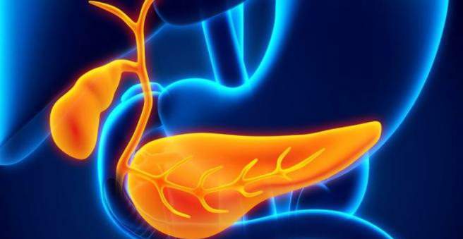
The liver produces 600 to 800 milliliters of bile daily (bile). It excretes various substances such as bilirubin, a breakdown product of the blood pigment hemoglobin. The substances enter the bile ducts with bile. These begin as the smallest bile capillaries between the liver cells and then flow together to form larger bile ducts. They combine to form a right and left liver passage. This results in the common hepatic duct (common hepatic duct). From him a passage goes off to the gallbladder (cystic duct). It then proceeds as a “ductus choledochus” to the duodenum, where it joins the pancreatic duct (pancreatic duct).
The bile is first transported by the liver into the gallbladder, where it is thickened and stored. Then it is released into the duodenum and with it all the substances that the body no longer needs.
Cholangiocellular carcinoma: symptoms
Cholangiocellular carcinoma often causes no symptoms for a long time. Therefore, the bile duct tumor is often recognized only at an advanced stage. Symptoms of patients with bile duct carcinoma include:
- Jaundice (jaundice)
- enlarged gallbladder without pain (palpable or visible in abdominal ultrasound)
These two symptoms are also summarized as Courvoisier signs. In addition, cholangiocellular carcinoma can cause the following symptoms:
- Stuhlentfärbung
- dark urine
- Itching (pruritus)
- weight loss
- Pain in the upper abdomen
- nausea
- Vomit
Cholangiocellular carcinoma: causes and risk factors
The exact cause of cholangiocellular carcinoma is unknown. There are several diseases that promote the development of bile duct cancer. These include:
- Extensions of the bile ducts outside the liver (choledochal cysts)
- Bile duct stones (choledocholithiasis)
- Parasitic diseases of the biliary tract (eg trematodes or liver fluke)
- Primary sclerosing cholangitis (also “PSC”, an inflammatory disease of the biliary tract)
Cholangiocellular carcinoma: examinations and diagnosis
From a bile duct tumor, the doctor must differentiate other diseases of the internal organs, such as a pancreatic carcinoma, which causes similar symptoms. Therefore, when suspecting a cholangiocellular carcinoma, he will first ask you about your medical history (anamnesis) and ask you questions such as:
- Have you lost weight inadvertently lately?
- Does it itching your skin?
- Is your stool lighter than usual, or is your urine darker?
- Do you have to vomit more often?
Physical examination
The doctor then examines her physically. Among other things, he scans your belly. He may feel an enlarged gallbladder beneath the last right rib in advanced bile duct cancer. If he comes along with jaundice, one calls the Courvoisier sign. It indicates a closure of the draining bile ducts. This has the consequence that the bile back up into the liver.
laboratory tests
The patient is also bled for suspected cholangiocellular carcinoma. This is being tested in the laboratory for specific values that might suggest bile duct carcinoma. These include the liver enzymes alanine aminotransferase (ALAT), aspartate aminotransferase (ASAT), glutamate dehydrogenase (GLDH), gamma-glutamyltransferase (γ-GT) and alkaline phosphatase (AP). They all may be elevated in liver damage. In addition, the bilirubin level in the blood is determined. The degradation product of the blood pigment leads to jaundice, among other things, if it is not sufficiently excreted via the bile.
Further diagnostics
A cholangiocellular carcinoma is best detected by ultrasound (sonography). It can happen that a cholangiocellular carcinoma is detected in a routine ultrasound examination of the abdomen.
In addition, endoscopic retrograde cholangiography (ERC) is often used for diagnosis. An endoscope, ie a tube with a camera at the front end, is advanced over the mouth and esophagus to the duodenum. There, the estuary of the ductus choledochus is visited and contrast agents are injected. Now an x-ray of the abdomen is made, on which the contrast medium can be seen. It should spread in the bile ducts. If it saves a bile duct, this is for example an indication of a stone or a tumor.
As an alternative to ERC, there is percutaneous transhepatic cholangiography (PTC). Contrast agents are also injected into the bile ducts. In this case, a needle, which is advanced under the X-ray control from the outside through the skin and the liver to the bile ducts.
An ERC or PTC may also be used for endosonography. This is an ultrasound scan in which the ultrasound head is not held on the skin, but placed in the body of the patient. In so-called intraductal sonography (IDUS), the access paths of the ERC or PTC are used to transport the miniscan heads into the bile ducts.
A cholangiocellular carcinoma can also be diagnosed using magnetic resonance imaging (MRI) or computed tomography (CT).
Cholangiocellular carcinoma: treatment
A cholangiocellular carcinoma is usually operated on. An attempt is made to remove the entire bile duct tumor. Depending on where it is located and how far it has spread, not only bile ducts but also the gallbladder and parts of the liver are removed.
If surgery is not possible or not successful, there are palliative treatment options. Palliative means that the patient can no longer be cured, but his symptoms should be improved by the therapy. For this you can use a so-called stent in the bile ducts. It is a small tube that keeps the bile ducts open, so that the bile can drain better.
In addition, one can try to keep the bile ducts open by means of radio frequency or laser therapy. The cancer cells are destroyed by heat. Chemotherapy with the active ingredients gemcitabine and cisplatin can also be used in palliative therapy.
Cholangiocellular carcinoma: disease course and prognosis
A cholangiocellular carcinoma has a poor chance of recovery. This is mainly because it is recognized late in many cases. The literature states that only two to 15 percent of patients still live five years after diagnosis.
The chance of survival depends primarily on whether the bile duct carcinoma could be completely removed in an operation or not. After a successful complete removal, up to 40 percent of the patients still live after five years. Should be cholangiocellular carcinoma not completely cut out, the survival rate is very low.