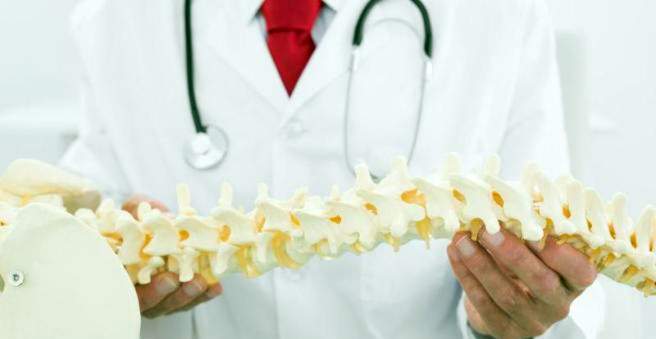
A vertebral fracture is caused by an indirect trauma (such as a fall) or osteoporosis. He can express himself through movement-related pain. Sometimes, however, a broken vertebra does not produce any symptoms and then often goes undetected. Depending on the fracture shape, a vertebral fracture can be treated conservatively or surgically. Find out more about the vertebral fracture here.

Vertebral fracture: description
The spine consists of a total of seven cervical, twelve thoracic, five lumbar, five fused cruciate and four to five coccyx vertebrae. Together with a complex ligament and muscle apparatus as well as the intervertebral discs and their characteristic double S-shape, the spine is a functional elastic system that can absorb loads.
The vertebral bodies together form the spinal canal, in which the spinal cord (part of the central nervous system) runs with all its orbits. From the spinal cord go the so-called spinal nerves (peripheral nervous system), which emerge laterally between the vertebrae.
If overloaded, the muscle-ligament apparatus may rupture and / or cause a vertebral fracture. This can hurt the spinal cord and spinal nerves.
Various forms of vertebral fracture
A vertebra consists of a vertebral body, the spinous process and the two transverse processes. Therefore, the vertebral fracture is subdivided into:
- Vertebral fracture
- Spinous process fracture
- Transverse process fracture
One differentiates between three different fracture forms, which can run in different directions (classification of Wolter and Magerl):
- Type A – Compression injuries: Here the vortex is compressed. This happens especially in the front of the vortex.
- Type B – Distraction injuries: A torque causes the vortex to break in the transverse direction. Such injuries occur in the posterior vertebral area.
- Type C – Rotation injuries: They occur during a rotation. Even longitudinal ligaments and not infrequently intervertebral discs are affected.
Vertebral fractures are additionally divided into stable and unstable fractures. This is important for the later therapy decision.
Stable vertebral fracture
In a stable vertebral fracture, the soft tissues as the surrounding ligaments remain undamaged, so that the spinal canal is not restricted and no neurological symptoms occur. The affected person can usually be treated early and mobilized.
For example, a stable vertebral fracture is a simple axial compression fracture (Type A). By compression of the vertebral body is stable against axial forces and also against forces in the diffraction direction. 85 percent of all spinal injuries are primarily stable fractures. The following vertebral body fractures are among the stable fractures:
- Isolated disc injuries
- Isolated vertebral fracture without disc injury, compression fractures
- Isolated vertebral arch fracture
- Vertebral fracture with disc injury
Unstable vertebral fracture
An unstable vertebral fracture occurs when the affected spinal column can be deformed by forces acting from different directions. These include, for example, distraction injuries (type B) and rotational injuries (type C). Once the posterior wall of the vertebral body is affected, one speaks of an unstable vertebral fracture, as there is a risk that the spinal cord is injured by displaced bone fragments. The injury can lead to paraplegia.
In case of unstable fractures, the affected person is limited in his mobility longer. The following vertebral fractures are unstable:
- Vertebrate fracture (mostly on the cervical spine)
- Rubble fracture with disc tissue damage and displaced fragments fore and aft
- Cracking fractures with a kink from 25 degrees
- Fractures of the articular processes with gaping spinous processes
- Vertebral arch injury
Vertebral fracture: symptoms
When a vertebra is fractured, local pain typically occurs – whether the patient is resting, moving, or performing stressful movements. Pain sufferers usually take a restraint. As a result, the surrounding muscles can tense up (muscle tension). In cervical vertebrae sufferers often support the head with his hands because of unstable head posture. Eventually, a bruise appears on the back of the neck.
If the vertebral fracture is accompanied by nerve damage, attacks of intense, severe pain (neuropathic pain) and painful burning or stinging (neurogenic pain) can occur. Emotional palsy (paresthesia) is also possible and mobility may be limited in the segment corresponding to the level of injury.
Vertebral fracture: causes and risk factors
A vertebral fracture can have different causes. They can be divided into two groups:
Traumatic conditional vertebral fracture
A vertebral fracture arises predominantly by an indirect force, for example, in a fall from high altitude on the legs (chain fracture), on the buttocks or the head. Direct traumas such as a spinal blow or an open vertebral fracture after a gunshot wound are extremely rare. But even with simple Bagatelltraumen such as a somersault on the exercise mat or a Stutz in the parking lot can lead to a severe spinal fracture with serious consequences.
In general, the transitions between the cervical spine and thoracic spine, between the thoracic spine and the lumbar spine and between the lumbar spine and the sacrum are particularly prone to injury. About half of all vertebral fractures involve the transition between the thoracic spine and the lumbar spine. The following typical situations can lead to trauma to the spine:
- Lap-belt injury (“seat belt injuries”) can cause a vertebral fracture along with abdominal injuries.
- When falling from great heights, a heel bone fracture often occurs along with a fracture of the thoracic and lumbar spine.
- Intervertebral disc and ligamentous structures can rupture if rapid body movement is stopped abruptly (deceleration trauma).
Spontaneous vertebral fracture
If a vertebral fracture develops without a corresponding accident, other causes must be considered. Especially in the elderly, osteoporosis (bone loss) plays a major role. The bone loses its bone mass and becomes unstable. Even a small force can then lead to a vertebral fracture.
A vertebral fracture caused by osteoporosis is also known as “sintering fracture”. Here, the base and cover plates break as a so-called fish vertebra or the front wall of the vertebral body as a so-called Keilwirbel. This is especially common in the lower thoracic spine and the upper lumbar spine. The dens fracture arises, for example, often in old people by a fall on the face (Dens = thorn-like extension of the second cervical vertebra).
Furthermore, bone metastases, bone tumors, ankylosing spondylitis, a plasmocytoma (multiple myeloma – a cancerous disease) and spondylitis (vertebral body inflammation) can lead to an unexpected vertebral fracture with slight deprivation.
Vertebral fracture: examinations and diagnosis
If you suspect a vertebral fracture, you should consult a doctor for orthopedics and trauma surgery. He will first ask you about a previous accident and your medical history:
- Did you have an accident? What happened?
- Was there a direct or indirect trauma?
- Do you have pain? If so, in which area and with which movements?
- Were there any previous injuries or previous damage?
- Have you had any complaints before?
- Do you have numbness on your arms or legs?
- Are there additional gastrointestinal complaints, difficulty urinating or dysphagia?
About ten percent of all spinal injuries also have nerve injuries. In addition, the doctor will think of a broken vertebra and the possibility of concomitant injuries. Usually a severe trauma is underlying, so kidney and spleen, for example, can be affected.
Clinical examination
In the clinical examination, the doctor checks whether walking or standing is possible. He also tests the general mobility of the patient. Next cranial nerves, sensibility and motor skills are examined to see if any neurological deficits are present. In addition, the doctor checks whether there is tension or hardening in the muscle (muscle harness) or a torticollis (torticollis).
Imaging procedures
An X-ray examination in two levels is an important part of the diagnosis of the vertebral fracture. Furthermore, functional recordings are made to accurately assess whether discs or ligaments have been injured. In addition, the distances of the spinous processes of the vertebrae, the vertebral body cavities and the vortex form are assessed.
For poorly visible areas, computed tomography (CT) is particularly well suited as an imaging procedure. This is especially true for the transition region of the cervical spine to the thoracic spine. Injuries in this area can be accurately estimated using CT. If there are nerve failures, a CT is always done.
Magnetic resonance imaging (MRI) is usually not required for acute injuries. It is only used to exclude spinal cord and disc injuries.
Vertebral fracture: treatment
In principle, the vertebral fracture therapy can be done both conservatively and surgically. Which method is best for each case depends on the type of injury (such as stable or unstable fracture) and also on the age of the patient.
Vertebral treatment: Conservative
If it is a stable fraction, it is usually treated conservatively. The patient is recommended to take care of themselves and to keep bed rest until the pain has improved. However, it can happen in some cases that the vertebral column can curve due to the altered shape of the fractured vertebral body. A strong curvature can lead to permanent discomfort. With a curvature from 20 degrees in the thoracic and lumbar spine area is therefore usually operated.
With a stable fracture of the cervical spine, this can optionally be re-aligned with an extension (Crutchfield) together with an X-ray control – the vertebral joints are thereby stretched in the axial direction. Then the cervical spine is immobilized with a soft collar (Schanz tie), a hard collar (Philadelphia tie), a Minerva plaster or a Halo fixator.
For the conservative treatment of the thoracic and lumbar spine can be treated with a three-point corset or a gypsum (plastic) corset.
Vertebral treatment: Operative
An unstable vertebral fracture is usually operated because there is always the risk that the spinal cord is injured or already injured. If the nerves are affected, a so-called laminectomy is performed, whereby parts of one or more vertebral bodies are removed.
The goal of surgical treatment is to quickly reorient and stabilize the spine to relieve pressure on the nerves as quickly as possible. This also applies to complete paraplegia, even if it can not be estimated whether an improvement will occur after surgery – it is always difficult to predict to what extent the spinal cord is damaged.
Spontaneous fractures, such as those caused by osteoporosis, involve either kyphoplasty or vertebroplasty. In traumatic fractures, two methods are used in principle: osteosynthesis or spondylodesis.
Vertebral surgery: kyphoplasty
Kyphoplasty is a minimally invasive method in which the broken vertebral body is re-erected with a balloon. Subsequently, the height of the vortex is stabilized by injecting cement.
Vertebral surgery: vertebroplasty
Vertebroplasty is also a minimally invasive method to stabilize the fractured vertebral body. Again, cement is injected into the vertebral body.
Vertebral surgery: osteosynthesis
In osteosynthesis, the bone fracture is screwed or flattened. A rupture of the dens (thorn-like extension of the second cervical vertebra) or a bilateral rupture of the vertebral arch is usually screwed. Fractures of the thoracic and lumbar spine are fixed over several segments (internal fixator).
Vertebral Surgery: Spinal fusion
In spondylodesis (stiffening surgery), two or more vertebrae are stiffened with a bone chip or plate. This procedure is usually considered for injuries of the ligaments and discs of the cervical spine. Plates are attached to the cervical spine from the front and from behind.
If the spine is bent over 20 degrees forward by a compression fracture in the thoracic and lumbar spine area, the vertebral fracture is fused from the front and the back. Distraction and torsion injuries of the thoracic and lumbar spine are also stiffened from both sides.
Vertebral fracture: Disease course and prognosis
Disease and prognosis in a vertebral fracture are usually good. However, it plays a big role, whether nerve tissue was injured. In addition, even after the trauma there is still the danger that the spinal canal is narrowed or neighboring segments change degeneratively. The following sequelae can occur after spine injuries:
static interference
Once the vertebral fracture has healed, there may be orthopedic problems with regard to statics.
spinal cord
All vertebrae are at risk of spinal cord or nerve root injury. In the extreme case, paraplegia occurs.
Post-traumatic kyphosis
If the vertebrae break in from the front, the posterior convex deflection of the spine may increase. In the thoracic spine, the bulge in the chest can multiply (“widow’s hump”) and diminish in the lumbar spine area.
Posttraumatic scoliosis
A lateral curvature of the spine (scoliosis) arises by lowering the side edges. This scoliosis is short-curved. The statics are influenced by the trunk overhanging and the overlying and underlying discs are increasingly stressed.
Schipperkrankheit
In severe physical work such as “shaking,” spinous processes can break from vertebrae, especially from the seventh cervical or first thoracic vertebra. This does not cause any significant complaints.
Vertebral fracture: healing time
The healing time in a vertebral fracture depends on how severe the injuries are. A stable vertebral fracture is usually bony again in a few weeks to months without moving further. Affected individuals may get up immediately or after about three weeks, depending on the pain. An unstable one vertebral fracture However, it may shift further, thereby increasing the risk of compression of the spinal cord and resulting in paraplegia.