Leishmaniasis (Leishmaniasis, Orientbane, Kala-Azar) is a tropical infectious disease caused by parasites called Leishmania. The Leishmania infection occurs worldwide in humans and animals and is transmitted by the sandfly. Leishmaniasis occurs in different variants. Read more about signs and treatment of leishmaniasis here.

Leishmaniasis: description
Leishmania are single-celled parasites and are transmitted via the saliva of the sandfly (butterfly mosquito). There are a total of 30 different Leishmania species, of which ten are pathogenic in humans and are distinguished by their appearance and clinical picture. Many mammals, especially rodents, serve as a natural host.
Leishmaniasis: human
Depending on the form of the disease, leishmaniasis causes skin ulcers in humans, affects the mucous membranes in the nasopharyngeal region or causes severe damage to the liver, spleen or bone marrow. Around 12 million people worldwide suffer from leishmaniasis, and two million become infected each year.
Leishmaniasis is particularly prevalent in the tropics and the Mediterranean. In Germany, leishmaniasis is rare, but fall ill – favored by mass tourism – vacationers again and again.
Leishmaniasis: forms
All forms are transmitted by different species of sandfly:
|
Leishmaniasis form |
befalls |
Leishmania species |
|
Cutaneous leishmaniasis of the “Old World” (Oriental bulge) |
skin |
Leishmania tropica |
|
Cutaneous leishmaniasis of the “New World” |
skin |
Leishmania vianna |
|
Cutaneous and mucocutaneous leishmaniasis of the “New World” |
Skin and mucous membrane |
Leishmania brasiliensis |
|
Visceral Leishmaniasis (Kala-Azar) |
Skin and internal organs |
Leishmania donovani |
Leishmaniasis: occurrence
The leishmaniasis of the “Old World” occurs about 90 percent in Europe and Asia. These include Afghanistan, Algeria, Saudi Arabia, Iran, Iraq, Ethiopia, the Middle East and the Spanish Mediterranean islands. The pathogens are mainly Leishmania tropica, Leishmania major and Leishmania aethiopica.
About ten percent of new cases are caused by infections in the “New World”. These include Central and South American countries such as Brazil, Mexico, Bolivia and Peru. The pathogens are predominantly Leishmania mexicana and Leishmania brasiliensis.
Leishmaniasis: symptoms
Long-lasting nodules or altered skin on the face or arms should always be thought of as leishmaniasis after traveling in high-risk areas. Often the symptoms of leishmaniasis are confused with those of a lymphoma (disease of cells of the lymphatic system).
Leishmaniasis symptoms: human
Symptoms can vary greatly depending on the type of leishmaniasis and how strong the patient’s immune system is. Immunity is always built only against the corresponding leishmaniasis species that has infected the host.
Cutaneous leishmaniasis of the “Old World”
In the case of cutaneous leishmaniasis of the “Old World”, a distinction is made in principle between three forms. Acute cutaneous leishmaniasis, Leishmania recidivans and disseminated cutaneous cutaneous leishmaniasis:
Acute cutaneous leishmaniasis
In acute cutaneous leishmaniasis, a small reddish brown nodule (papule) usually develops and enlarges rapidly. The altered area of the skin has a raised edge that can easily become infected. The wound heals after about a year and usually leaves behind an ugly pigmented scar. Especially cheeks and arms are affected.
The acute cutaneous leishmaniasis can be divided into three other types, which are each transmitted by different species of sandfly.
- Dry or urban leishmaniasis (Leishmania tropica): The classic form of leishmaniasis, which causes the skin damage described above.
- Moist or rural leishmaniasis (Leishmania major): Sudden infection with skin damage.
- Ethiopian Leishmaniasis (Leishmania aethiopica): Less severe leishmaniasis, however, can become chronic and cause diffuse damage to the skin.
Leishmania recidivans
Leishmania recidivans is a form of dry or wet leishmaniasis and is chronic. The skin damage is initially soft and heals from the middle. In addition, serpentine brown-reddish nodes develop near older nodes.
Disseminated diffuse cutaneous leishmaniasis
Disseminated cutaneous cutaneous leishmaniasis is a rare variant in immunocompromised patients. The infection spreads out to form countless papules and nodules on the skin and mucous membranes. It is also reminiscent of leprosy (leprosy lepromatosa).
Cutaneous leishmaniasis of the “New World”
The multiple symptoms vary, cause major skin damage and are more chronic than cutaneous leishmaniasis of the “Old World”.
Leishmaniasis of the “New World” is also known as:
- Chiclero Ulcer (Gum Ulcer): A form that affects the ear cartilage. In particular, forest workers are affected.
- Uta: A variant that occurs in the Andean Highlands. Occasional skin damage occurs.
Cutaneous and mucocutaneous leishmaniasis of the “New World”
After a first infection in the face, the pathogen spreads via the blood or the lymphatic system to the mucosa of the nasopharynx. After months to years, the nose and lip area thicken and the pathogens destroy the nasal septum. Initially, the anterior cartilaginous septum is usually affected. Typical symptoms are a stuffy nose. Affected are the throat, the uvula, palate and tongue to the larynx and trachea. Mucocutaneous leishmaniasis can be chronic and lead to sepsis.
Visceral Leishmaniasis (Kala-Azar)
Visceral leishmaniasis is the most dangerous form and can affect the entire organ system of the body. In addition to the skin, liver, spleen, bone marrow and lymph nodes are also affected. The incubation period ranges from three months to two years. Patients repeatedly suffer from high fever, chills, weakness, anemia, skin and mucosal bleeding and hair loss. The skin develops small, dark red papules, brownish-yellow raised spots and discolors grayish in the course of the illness. If visceral leishmaniasis is not treated, the “black disease” will be fatal within one to two years.
Surviving patients may develop Post-Kala-Azar skin leishmaniasis after one to three years. In the face or on the body, bright or reddish spots appear, which change into papules and nodules. The appearance is often reminiscent of the leprosy disease (leprosy lepromatosa).
Leishmaniasis: causes and risk factors
The infectious disease leishmaniasis is caused by parasites of the genus Leishmania (protozoa) that live in humans or in animals. Even pets, like dogs, serve as hosts. The pathogens are particularly easily introduced when, for example, dogs from the Mediterranean are introduced.
Leishmaniasis: contagion
The sandfly is a vector (vector) of leishmaniasis, in particular sandflies of the genera Phlebotoma, Lutzomya and Psychodopygus. It is only a few millimeters in size and especially in warmer areas. If she stings an infected host, she picks up the parasites. The Leishmania develop further in the body of the sandfly and are transferred to a new host on the next trick.
The disease can also be transmitted via blood transfusions, bone marrow or organ transplants. Rarely does the disease pass from mother to child during pregnancy.
Only in 2014, a sandfly species was discovered in Hesse. Whether it can also infect people with Leishmaniasis, has not been proven. Leishmaniasis occurs at any age.
Cutaneous leishmaniasis of the “Old World”
The Skin Leishmaniasis of the “Old World” is also called Baghdad Bump, Aleppo Bump, Jericho Bump or Orient Bump. It occurs mainly in the Mediterranean, the Middle East and the Sahara highlands and is caused by Leishmania tropica. Their natural hosts are infected rodents and humans. The incubation period for an infection with Leishmania tropica ranges from a few days to several years – usually two to four weeks.
Cutaneous leishmaniasis of the “New World”
The cause of skin-leishmaniasis of the “New World” is an infection with the species Leishmania mexicana or Leishmania vianna.
Cutaneous and mucocutaneous leishmaniasis of the “New World”
Mucocutaneous leishmaniasis is known in Argentina and Peru as “Espundia” and as “Bouba” in Brazil. Pathogens are parasites from the Leishmania vianna complex. Leishmania brasiliensis is the most common form. This pathogen complex occurs mainly in the jungle areas of Brazil and Peru.
Visceral Leishmaniasis
Visceral leishmaniasis is also called black fever, Dumdum fever or Kala-Azar (“black death”). It occurs mainly in India, South America, China, Sudan and other African regions as well as in the Mediterranean countries. Pathogens are Leishmania from the Leishmania donovani complex.
Leishmaniasis: examinations and diagnosis
If you have any suspected symptoms, you should consult a specialist in dermatology, infectiology or tropical medicine. Diagnosis is based on the symptoms, the medical history (anamnesis) and the microbiological detection in the laboratory. For example, the doctor will ask you the following questions:
- Did you have a fever? If so, how did the fever go?
- Have you been traveling in recent years? In which countries?
- Do you suffer from other comorbidities with weakened immune systems, such as HIV infection?
Even though your travels to southern countries, the tropics or subtropics have been a long time ago, you should alert your doctor.
Leishmaniasis: further diagnostics
The doctor takes a small tissue sample from the edge of the affected skin. In the laboratory, the Leishmania, which are about two to five thousandths of a millimeter in size, are stained in this sample with a special dye (Giemsa stain) and thus made visible under the microscope. Also via a so-called culture, the pathogens can be discovered by growing the parasites in a nutrient medium. The exact species can be determined with further laboratory tests.
The Montenegro reaction is an immunological test in which heat-killed Leishmania are injected under the skin. In the case of an infection, a vaccination papule of one to two centimeters in diameter develops on the reddened skin after 48 hours. Even long-standing leishmaniasis infections can usually still be detected.
Old scars may indicate cutaneous and mucocutaneous leishmaniasis of the “New World” – as well as an increased number of antibodies in the blood and a positive skin test. For the detection of visceral leishmaniasis, the doctor takes a sample of bone marrow, spleen or liver. Also in the blood, the pathogens can usually be detected. In patients with weakened immune systems, this evidence is not always possible.
Post-Kala-Azar skin leishmaniasis can be detected via a tissue sample of the skin.
Leishmaniasis: treatment
The acute cutaneous leishmaniasis of the “Old World” usually heals spontaneously and requires no therapy. Often it is already treated successfully by icing or surgical procedures (electrocoagulation). For more stubborn cases, the doctor injected an antimicrobial locally into the skin. The treatment is continued one to two times a week for two to four weeks.
The doctor treats leishmaniasis of the “Old World” mostly locally. Visceral leishmaniasis, as well as the cutaneous and mucocutaneous form of the “New World”, require systemic, ie whole-body, therapy, and infusion of amphotericin B or antimony supplements.
Various medicines are available for leishmaniasis therapy:
Antimony supplements: Antimony is a heavy metal that inhibits Leishmania’s metabolism. It is generally used for infections with Leishmania brasiliensis and visceral leishmaniasis.
Pentamidine: Pentamidine is mainly used in diffuse cutaneous leishmaniasis and possibly in the mucocutaneous form.
Amphotericin B: This medication is normally used against fungi, but also works against protozoa. If antimony supplements cause side effects or are not effective enough, amphotericin B is an alternative in visceral leishmaniasis.
Ketoconal: Ketoconal, an antifungal agent, can be used by the doctor for infections caused by Leishmania mexicana and Leishmania major.
Leishmaniasis: disease course and prognosis
Cutaneous leishmaniasis is the easiest compared to other leishmaniasis diseases, but there is always a scar. Mucocutaneous leishmaniasis is more severe as it primarily damages the nasopharynx and must always be treated systemically. The same applies to visceral leishmaniasis, which can be fatal.
Leishmaniasis: prevention
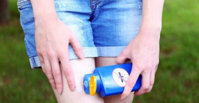
There is no leishmaniasis vaccine yet. In risk areas, you should first and foremost be aware of the sandfly and thus of infection Leishmaniasis pathogens protect. A raised sleeping space and impregnated bed nets provide protection during the night. Also remember to bring adequate mosquito repellent and long clothes. For dogs there are special collars.