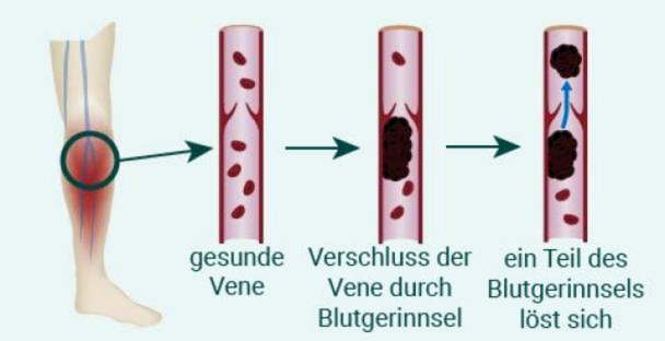
Thrombosis is a vascular occlusion by a blood clot. Most commonly, thrombosis occurs in the veins in the leg. Important signs of thrombosis include swelling, pain and a red or bluish discoloration of the skin. Even fever is possible. Thrombosis is dangerous because the clot can dissolve and flush into other organs. Read all important information about thrombosis symptoms, treatment and prevention!

Thrombosis: short overview
- Most common locations: Legs (especially lower leg), pelvic area, arms, upper or lower vena cava
- Typical symptoms: Swelling, redness, overheating, pain and distension, fever, accelerated pulse
- Treatment: Compression bandage or compression stockings, elevated storage, anticoagulant drugs, thrombectomy (OP)
- Important investigations: Ultrasound image, venography (“vein X-ray”), CT, blood test (D-dimers, coagulation factors)
- Hazards: Pulmonary embolism (pulmonary artery occlusion), vascular and tissue damage (postthrombotic syndrome)
- Special form: Anal thrombosis (anal venous thrombosis)
Thrombosis symptoms
There are a number of very typical signs of thrombosis. Depending on where the clot has formed, the symptoms are partially different.
Symptoms of thrombosis in the leg
Thrombosis is particularly common in the large veins of the lower leg. Because there, the blood flows against the gravity especially slowly back to the heart. The most common signs of thrombosis are then:
- Swelling of the calf, often the ankle region and the foot
- Severity and tension in the lower leg
- Pain in the lower leg, sometimes in the foot, thigh or groin, which may resemble sore muscles
- tense (shiny) and bluish discolored skin
- Overheating of the lower leg
- more visible skin veins (so-called warning veins)
- light fever
- accelerated pulse
Thrombosis in the arms usually cause more discomfort than vascular occlusions in the legs. In addition, they are noticeable by an increased vein pattern, because the blood seeks its way to the heart via detours.
Even if some of these symptoms are missing, a thrombosis in the leg is nevertheless not excluded. Neither are the said thrombosis signs proof that there really is a leg vein thrombosis.
Symptoms of thrombosis in the arm
The veins in the arm can be closed by blood clots. Overall, this is much less common than in the leg. Typical thrombosis symptoms in the arm are:
- Swelling and overheating of the affected arm
- Swelling of the hand
- bluish skin veins
- partially reddish-purple discoloration of the arm
- Pain with pressure on the arm and when moving
Basically, thrombosis can occur in all blood vessels of the body. Unlike thrombosis in the extremities, the symptoms are often ambiguous. There may be severe pain or dysfunction of organs. To clarify such unspecific symptoms of thrombosis more and more medical examinations are required.
Symptoms of anal thrombosis (anal venous thrombosis)
An anal thrombosis is noticeable by a painful swelling in the anal area. The anal thrombosis is often difficult to distinguish from the so-called hemorrhoids. But it has another cause: in the case of anal thrombosis, the blood clot is formed in a small vein of the lower anal canal. The painful hemorrhoid, on the other hand, is an enlarged blood vessel of an arteriovenous vascular pad that penetrates outward from the inner anal canal.
Anal venous thrombosis is very painful, especially because it lies directly in the area of the opening. The anal thrombosis, however, can usually be treated well. Here you can learn more about the symptoms, causes and therapy of anal thrombosis!
Thrombosis Treatment
A thrombosis can be treated by medication, by compression therapy or surgically. Which method is used, inter alia, depends on the location where the clot has formed. Often, however, the different treatment approaches have to be combined.
The most important goal of thrombosis treatment is to prevent the clot from detaching from the vein wall and trailing the bloodstream into vital organs. Because then there is the danger of a so-called embolism (for example, a pulmonary embolism), ie the clogging of an artery through the clot with potentially life-threatening consequences. In addition, long-term, irreparable damage to the affected blood vessels, extremities or organs (post-thrombotic syndrome) should be avoided.
Elevation and compression
Important immediate measures for a recent thrombosis in the extremities are to uplift the affected leg or arm and apply a compression bandage. This prevents the blood from damaging more and the extremity from swelling.
The compression bandage must extend well beyond the site of the thrombosis – in the case of a lower leg thrombosis, ie below the knee. He must be taut to squeeze the veins so much that the blood flows better inside them. He may not constrict the extremity at any point.
A good way to achieve a sufficiently strong and consistent degree of compression is compression class II thrombosis stockings. Compression treatment should be continued in the long term if veins have been damaged by the thrombosis.
Thrombosis treatment with drugs
The drug thrombosis treatment is intended to prevent the blood clot from growing and possibly being flushed into the pulmonary arteries. In the best case, the drug can cause the body’s own substances (enzymes) to reduce the thrombus again or even completely dissolve. Anticoagulant drugs can also prevent the re-emergence of thrombosis.
Acute treatment of thrombosis
One begins the treatment of thrombosis with a so-called initial anticoagulation, which should begin immediately if the diagnosis thrombosis was made or if thrombosis is very likely the cause of discomfort.
For this purpose, the drug is usually heparin, which inhibits blood clotting. Heparin must be given in high doses as a syringe under the skin (subcutaneous injection) or as an infusion. Because the drug would disintegrate in the gastrointestinal tract and then not get into the bloodstream.
The active ingredient fondaparinux is injected under the skin. It is mainly used when patients have previously reacted to a heparin dose with a life-threatening decrease in the number of blood coagulation platelets = platelets). Other active ingredients of the acute thrombosis treatment are the so-called DOACs (direct oral anticoagulants) rivaroxaban and apixaban.
Long-term treatment after a thrombosis
Afterwards, usually after about five to ten days, the patients receive an anticoagulant drug in tablet form to prevent a new clot from forming. This so-called maintenance therapy will continue for three to six months. For this purpose, so-called vitamin K antagonists are used. These are antagonists of important for blood clotting vitamin K. Especially the active ingredients phenprocoumone and warfarin are used in Germany.
Important: The correct dosage of these drugs must be regularly checked by blood tests of the so-called coagulation values!
Operative thrombosis treatment
Especially in young patients experiencing thrombosis in a large vein in the leg and pelvic region for the first time, surgical intervention may be the best treatment option. An attempt is made to grasp the blood plug (thrombus) using a catheter and to pull it out of the vein. Doctors also speak of “recanalization”, because with the procedure a blocked blood vessel is made through again. The doctors also check if there is a flow obstruction in the vein that can be eliminated.
Often, a thrombus-dissolving drug is given over the catheter. This localized form of thrombosis treatment has better success rates and lower risks than the systemic treatment that used to be common in the past, where the drug had to be distributed throughout the body in high doses.
Recanalization therapy should be performed as early as possible to reduce the risk of postthrombotic syndrome. Possible complications of this type of thrombosis therapy are bleeding, but also accidental detachment of clots. These could then continue in the vein path towards the heart and the pulmonary circulation.
In some cases, patients with leg vein thrombosis are implanted with a sort of “sieve” into the vena cava filter, either permanently or temporarily. It is intended to prevent detached blood clots from being flushed into the lungs. This procedure is suitable for patients who repeatedly suffer from pulmonary embolism despite anticoagulant drugs.
Thrombosis: causes and risk factors
Thromboses are blood clots that form in the blood vessels – almost always in veins. You can basically have three different causes, which may exist alone or in combination:
- Flow obstacles in the blood vessel: Damage / diseases or deposits on the vessel wall or constrictions of the blood vessels due to mechanical pressure from outside (for example scarring, tumors)
- slow flow rate: In abnormally dilated veins (varicose veins), by the effect of gravity or / and by a too low muscle tension (in case of immobility, paralysis or after operations), dehydration (blood becomes thicker)
- increased coagulation tendency of the blood: Diseases of the blood coagulation system, severe systemic diseases (cancer, autoimmune diseases), drug side effects (such as the “pill”), smoking
Traveling thrombosis and thrombosis after surgery
The blood backflow to the heart must work in the deep veins of the leg against gravity. This is supported in healthy, physically active people by two mechanisms:
- Venous valves: They act like valves and allow the blood to flow only in one direction, namely to the heart.
- Muscle Pump (Muscle Vein Pump): Through the work of the (calf) musculature, the veins in the leg are compressed again and again. In cooperation with the venous valves, the blood is thus pressed towards the heart.
If one or both mechanisms fail to function, blood flow can slow down greatly and the risk of thrombosis increases. This is the case, for example, when sitting in the car, the train or the plane for a long time. Therefore, one often speaks of a “traveling thrombosis”. But even sitting for hours on the computer can increase the risk of thrombosis.
Similarly, after injury or surgery, after which the leg is sedated or generally strict bed rest must be met, the natural effect of the muscle pump. Since every trauma – and surgery in the broader sense of the word – significantly increases the blood’s ability to coagulate, the risk of thrombosis after surgery is greatly increased.
Thrombosis in varicose veins
Varicose veins are highly dilated blood vessels. They occur particularly frequently in the area of the legs, in particular the lower leg. In varicose veins the blood flows more slowly and in addition, the natural valves in the veins, the venous valves, no longer function properly. This also increases the risk of thrombosis.
Thrombosis: diagnosis and examination
In a venous thrombosis in one leg, this is overheated and swollen. Certain pressure points and movements trigger pain, which the doctor (usually a specialist in internal medicine) can determine with a physical examination. Typical examples are:
- Calf pain when the toe is raised (Homans sign)
- Pain when pressing the calf (Meyer sign)
- Pressure pain on the inside of the foot (Payr sign)
In addition, an ultrasound examination can depict the occlusion of veins. In general: A superficial thrombosis is characterized by greater discomfort and is therefore often easier to diagnose than a vascular occlusion in deeper veins (phlebothrombosis). The latter, however, often has serious consequences.
With a phlebography (also: phlebography), the blood vessels in the body can be displayed on an X-ray. The procedure is therefore well suited for the diagnosis of deep vein thrombosis. For this purpose, a contrast agent is injected into a superficial vein on the back of the foot. To ensure that the contrast medium finds its way into the deep veins of the leg, the veins near the skin surface are first tied off with a moderately tight bandage. Where there is a thrombosis, the flow of the contrast agent is interrupted or “constricted”.
A much used examination procedure in vascular medicine is computed tomography (CT). In this method, the patient’s body is virtually sliced by X-rays. Due to the high image density can also be represented vessels and organs. This method is used for example in thrombosis in the abdomen or in a cavernous sinus thrombosis in the head.
In rare cases of vascular occlusions, such as thrombosis in the eye, a specialist ophthalmologist can make a picture of the retina and thus detect a stasis.
blood test
In addition to imaging, a blood test is important. This is looking for breakdown products of blood clots, the so-called D-dimers. It should be noted that the D-Dimer Thrombosis Test should only be used to exclude a blood clot if there is a high likelihood of vascular occlusion. A broad screening is not feasible with this blood test.
Thrombosis & pregnancy
If a thrombosis occurs during pregnancy or after a miscarriage, additional examinations should be performed to find the cause. As a result, a similar course in a later pregnancy may be avoided.
Other special cases
Also in thrombosis, which have no clear cause or occur in atypical vessels, the doctor will also try to find the cause of clot formation. For example, some people suffer from hereditary diseases that can interfere with blood clotting.
Thrombosis: disease course and prognosis
Thrombosis is a very serious illness and can lead to dangerous complications. These arise,
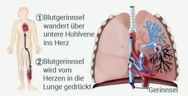
- when a blood clot dissolves and migrates to the heart, from where it can enter the lungs, for example (pulmonary embolism)
- if a vein can be blocked by a thrombus and permanently damaged (consequence: postthrombotic syndrome)
Pulmonary embolism is a particularly common and life-threatening complication of thrombosis. The thrombus (or parts of it) is flushed with the blood stream through the venous system to the right ventricle and from there into the pulmonary arteries. If he places a large artery there, a large part of the lungs will no longer be supplied with blood.
He can then no longer participate in the gas exchange, whereby a life-threatening oxygen deficiency can arise. In addition, the right ventricle can be excessively loaded by a high flow resistance; Right heart failure is also possible due to a pulmonary embolism. An embolism is therefore always a medical emergency!
Postthrombotic syndrome
Although the majority of thromboses heal without consequences, one-third of patients suffer from a so-called post-thrombotic syndrome. This causes varicose veins due to a blood drainage disorder, which persists even after reopening of the affected vessels. This drainage obstruction can lead to further tissue damage or reoccurrence of blood clots.
Prevent thrombosis
The best thrombosis prophylaxis (= prevention) is to avoid or reduce these thrombosis risk factors. For example, you should pay attention to sufficient exercise, especially on long air travel, but also during long office work days. In addition, a sufficient supply of liquid (drinking, liquid food) is important to keep the blood fluid and thus to avoid the formation of a clot.
Thrombosis syringes
After an injury or surgery or other illness-related immobilization, one can prevent clots with drugs: Daily thrombosis injections with heparin can prevent the formation of a blood clot in most cases.
Anti-thrombosis stockings
So-called anti-thrombosis stockings are special, elastic stockings made of a skin-friendly, thin fabric that either reach down to the knee, or even over the knee beyond the thigh. The light pressure that they exert on the veins causes the blood to flow back to the heart a little faster and more evenly.
In particular, when thrombosis risk factors such as a tendency to varicose veins exist, before and after surgery, or for long trips wearing anti-thrombosis stockings is recommended. Often they can help one thrombosis to prevent.
Additional information
Book recommendations:
- Guide to varicose veins, leg swelling and thrombosis (Erika Medoza, 2016, Springer-Verlag)
guidelines:
- S3 Guideline “Prophylaxis of Venous Thromboembolism (VTE)” of the Association of Scientific Medical Societies (as of 2015)
- S2k guideline “Diagnosis and Therapy of Venous Thrombosis and Pulmonary Embolism” of the German Society for Angiology – Society for Vascular Medicine (as of 2015)
Support Groups:
German Society for Angiology – Society for Vascular Medicine e.V.
House of the Federal Press Conference
Schiffbauerdamm 40
10117 Berlin
http://www.dga-gefaessmedizin.de/startseite.html
Action alliance thrombosis:
http://www.risiko-thrombose.de/
German Vascular League e.V.
Mühlenstr. 21-25
50321 Bruehl
http://www.deutsche-gefaessliga.de