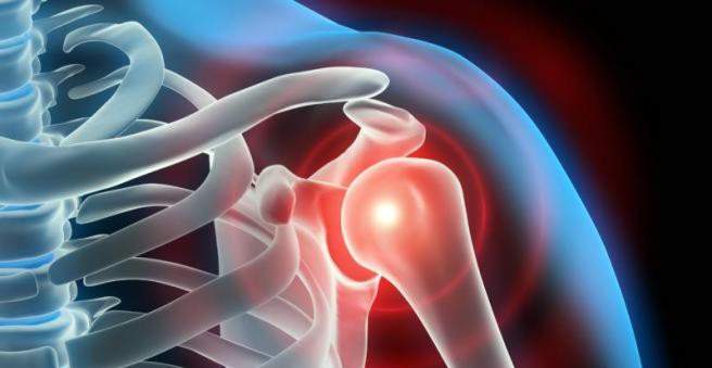
The impingement-shoulder syndrome (subacromial impingement syndrome) refers to a compression of muscles, tendons or nerves under the shoulder roof in the subacromial space. The result is dysfunction of the shoulder joint and pain. Protection, painkillers and physiotherapy can relieve the symptoms of impingement-shoulder syndrome. An operation can prevent permanent joint stiffening. Read more about impingement shoulder syndrome here.

Impingement Shoulder: Description
The impingement-shoulder syndrome is a painful compression of muscles, tendons or nerves in the area of the shoulder joint, more precisely in the subacromial space. This is the space between the shoulder roof (acromion) and the humeral head. Here the supraspinatus tendon runs, protected by a bursa (Bursa subacromialis). Four cuffed muscles surround the shoulder joint (rotator cuff). The tendons of the rotator cuff muscles can no longer slide freely through the compression in the joint space.
Two forms of impingement shoulder syndrome
The impingement-shoulder syndrome is divided into a primary outlet impingement syndrome and the secondary non-outlet impingement syndrome.
The primary Outlet Impingement Syndrome The shoulder is caused by a change in the bony structures. Degenerative structural changes or a bone spur can be the cause of a narrowing of the joint space.
The secondary Non-outlet impingement syndrome the shoulder, on the other hand, is based on a non-bony change. The inflammation of the bursa (bursitis) and muscle or tendon damage can reduce the joint space and provide for movement restrictions and pain.
Impingement-shoulder syndrome: frequency
In Germany, about ten percent of the population suffer from impingement-shoulder syndrome during their lifetime. Men and women are about the same age around the age of 50 affected. The shoulder joint is the most articulated ball joint in the body and has high elasticity, but at the same time makes the joint vulnerable to injury.
Impingement shoulder: symptoms
In the early stages, the impingement-shoulder syndrome is characterized by an acute onset of pain. At rest, he expresses himself only discreetly, but in stressful activities, especially when these are carried out over the head, he reinforces himself. In many cases, patients can identify a triggering event. Extraordinary stress in over-the-head activities or the influence of cold are often associated with the onset of pain. The pain in impingement-shoulder syndrome is referred to as lying deep in the joint. In addition, lying on the affected side is described as extremely unpleasant, as it increases the pain.
If the arm hangs loosely on the body and then lifted laterally in an extended posture (abduction), report patients with an impingement-shoulder syndrome from about 60 degrees of severe pain. An abduction between 60 and 120 degrees is impossible because the supraspinatus tendon is pinched. This phenomenon is described as a painful arc and is an important clinical sign of Impingement Shoulder Syndrome. Those affected often take a restraint and prevent painful movements. The inflammation of the bursa (Bursa acromialis) can cause adhesions and adhesions, which further aggravates the painful restriction of movement. In addition, defensive posture often leads to muscular atrophy due to the lack of exercise, which further reduces the stability of the shoulder joint.
Impingement Shoulder: Causes and Risk Factors
The Outlet Impingement Shoulder Syndrome results from the narrowing of the subacromial space due to bony changes in the shoulder, such as in the case of joint wear (osteoarthritis).
In non-outlet impingement shoulder syndrome, the surrounding soft tissues cause discomfort, such as bursitis. It is usually accompanied by swelling, which narrows the joint space. The supraspinatus tendon or biceps tendon may also inflame. Such tendonitis (tendinitis) also leads to a painful constriction of the joint space and resulting movement restrictions. In some cases, a tendon may also tear completely, causing the shoulder joint to loose a lot of stability (“instability impingement”).
Impingement shoulder: examinations and diagnosis
The right contact for suspected impingement-shoulder syndrome is a specialist in orthopedics and trauma surgery. He will first start the medical history (anamnesis) by asking you various questions, for example:
- Since when is the pain?
- Was there a severe strain or injury at the onset of pain?
- Does the pain increase during exercise, at night or when lying on the affected side?
- Do you suffer from movement restrictions in the affected joint?
- Does the pain radiate from the joint and is it of dull quality?
- Do you do sports and if yes, which ones?
- What do you do for a living?
Physical examination
The doctor will examine you physically after the first interview. He will test the mobility of the shoulder joint by asking the patient to lift his arm from his loosely hanging down position over his head and in an extended position. The painful arc is a typical clinical sign of the shoulder impingement syndrome (see above: Symptoms).
The degree of strength of the shoulder joint musculature is measured by the movement against resistance. There are several clinical tests that can be used to examine the individual muscles of the shoulder joint for damage. In addition, the doctor may ask the patient tonerve pinch“Exercise by placing both hands with the thumb pointing down the neck. At the “apron grip“The affected person is asked to grab behind his back with both hands as if he were putting on an apron. In an impingement-shoulder syndrome, patients complain of pain and can not comply with the requests.
Jobe test
The Jobe test is an orthopedic test used in clinical examination of the impingement syndrome (shoulder) to confirm or exclude involvement of the supraspinatus muscle and its tendon. For this, the patient is asked by the doctor to spread the arms at shoulder height (90 °) with the elbow extended and to turn the hands together with the forearms forward inwards (internal rotation). Now the patient should be able to withstand the pressure exerted on the upper arms by the doctor from above. If the patient is unable to hold his arms upright against resistance or reports pain, the test is positive and most likely there is supraspinatus damage. If the Jobe test is negative, look for other causes of the impingement syndrome (shoulder).
Impingement test according to Neer (Neer test)
Neer’s impingement test is another clinical trial for suspected impingement-shoulder syndrome. In doing so, the patient should extend his arm forward and screw in the hand and forearm maximally inwards (pronation position). The doctor fixes with one hand the shoulder blade of the patient and raises the arm of the patient with the other hand. The Neer test turns out to be positive when pain occurs when lifting the arm above 120 °.
Hawkins test
The Hawkins test is also a clinical test that can confirm or rule out impingement shoulder syndrome. However, it is far more unspecific than the Jobe and Neer tests. No individual muscles can be identified as the cause. The shoulder joint is passively rotated by the examiner in the Hawkins test. If pain occurs, the test is considered positive.
Impingement Shoulder Syndrome: Imaging
The impingement-shoulder syndrome can be detected using various forms of imaging. The X-ray examination is the first choice for detecting bony changes. An ultrasound examination (sonography) is used to detect any fluid accumulation in the joint space. Magnetic resonance imaging (MRI) is also used to visualize the surrounding soft tissue and fluid collections.
X-ray examination in impingement-shoulder syndrome
X-ray examination is the imaging diagnostic of choice to diagnose impingement shoulder syndrome. Bony changes can be detected and a joint overview created.
Ultrasound in impingement-shoulder syndrome
As part of an inflammation of the shoulder joint often occur fluid accumulation within the bursa. They can be easily and inexpensively detected by ultrasound (sonography). Sonography may also include other bursa changes, the muscular structures of the shoulder joint, and possible thinning of the muscles. All this provides evidence of a shoulder impingement syndrome.
Magnetic Resonance Imaging in Impingement Shoulder Syndrome
Magnetic Resonance Imaging (MRI) uses radio waves and magnetic fields to create very accurate images of muscles, tendons and bursae. It is superior here to the ultrasound examination, but also much more complex and cost-intensive. An MRI is particularly useful in upcoming joints reconstruction surgery to better assess operating conditions in advance.
Impingement Shoulder: Treatment
The impingement-shoulder syndrome is treated with various treatment approaches. The complaints are first tried to treat in a conservative way (physical protection, painkillers and physiotherapy). For complete healing, however, the impingement-shoulder syndrome must usually be operated on (causal therapy).
Conservative therapy
Conservative therapy includes first and foremost the protection of the shoulder joint and the avoidance of stress factors such as exercise or strenuous work over the head.
The drug treatment provides anti-inflammatory analgesics such as ibuprofen or acetylsalicylic acid. As a rule, however, they only relieve the symptoms and do not remedy the triggering cause.
A physiotherapeutic treatment aims to strengthen the surrounding muscles and specifically relieve the joint space of the shoulder joint. Physiotherapy teaches special impingement syndrome shoulder exercises, which the patient is free to do at home to relieve the symptoms.
Above all, the exercises serve to strengthen the muscle group of the shoulder joint, which is required for external joint rotation (external rotation): Targeted training of the so-called external rotators (rotator cuff) enlarges the joint space, which relieves strain.
In addition, because the muscles are dwindling with prolonged restraint (muscle atrophy), impingement shoulder exercises can also help to maintain the strength of the muscles. However, the affected shoulder joint should not be overloaded. Only a properly performed, regular physiotherapy can lead to decreased pain. Try to incorporate the learned exercises firmly into your daily routine in order to achieve the best possible therapeutic success.
Causal therapy
Causal therapy aims at impingement syndrome (shoulder) to treat the cause of the disease and remove it permanently. Structural changes can be removed by surgery (arthroscopy), which eliminates the mechanical tightness of the shoulder joint.
Arthroscopy (joint mirroring): Arthroscopy is a minimally invasive surgical technique in the articular area that is particularly recommended in young patients to minimize the risk of joint stiffness. Two to three small skin incisions are used to introduce a camera with an integrated light source and special surgical devices into the joint. The doctor can thus examine the joint from the inside and get a precise overview of the causative changes. Subsequently, he can expose the joint space, for example by abrading an existing bone spur or eliminating possible cartilage damage. If the impingement-shoulder syndrome has already caused tendon tears in the advanced stage, these can be sutured in the context of arthroscopy. The skin incisions only require fewer sutures for closure and leave only very subtle scars compared to open surgery.
Impingement shoulder: disease course and prognosis
The prognosis in the impingement-shoulder syndrome can not be generalized, since it depends on the triggering cause. In many cases, physiotherapy treatment must be performed over a long period of time before satisfactory results are achieved. The symptoms can be relieved in most cases by anti-inflammatory analgesics (anti-inflammatory drugs). However, this is not a permanent solution.
In many cases, the impingement syndrome (shoulder) leads to signs of wear and inflammatory reactions in cases of pronounced narrowing of the joint space. Persistent constriction can lead to compression of nerves and tendons as well as tissue rupture and death (necrosis). The risk of joint stiffness increases with increasing posture. Since even after surgery by the patient often automatically a restraint is taken, are subsequently always physiotherapeutic Impingement ShoulderExercises recommended.