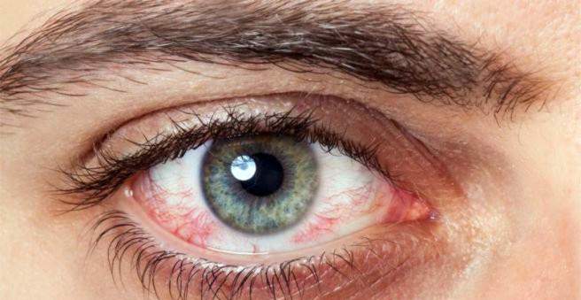
In corneal inflammation (keratitis) inflames the transparent part of the outer eye skin, which leads to severe pain. The triggers are mostly bacteria, viruses or parasites, but there are also no infectious causes. Advanced keratitis can lead to permanent vision damage. Read here the most important information about corneal inflammation.

Corneal inflammation: description
The eye can develop various inflammations – both outside and inside the organ of vision. Depending on which structures are affected, one must expect some dangerous complications. In the case of corneal inflammation (keratitis), a very important part of the eye is inflamed with the cornea. Therefore, special care should be taken with this condition.
What is the cornea and what is its function?
When you look at a human eye from the outside, the cornea (medical: cornea) initially does not show up, because it is transparent. It sits in the middle of the eyeball and forms the anterior surface in front of the pupil and the iris. If the pupil is the window of the eye through which the light rays are incident, then the cornea is, so to speak, the window glass. This also makes it clear why visual acuity may be impaired in corneal inflammation.
Around the cornea, the dermis (sclera) follows, which gives the eye white its color. The border between cornea and sclera is called limbus.
The cornea protects and stabilizes the eye, on the other hand, it has a refractive effect. Together with the lens, it is responsible for bundling the incoming rays of light into a focus on the retina. Without a cornea, no sharp vision would be possible.
How is the cornea structured?
The cornea is slightly smaller than a 1-cent piece and evenly arched. It consists of several layers, including from outside to inside:
- Epithelial layer: absorbs nutrients from the tear film and absorbs oxygen;
- Stroma: gives the cornea hardness and elasticity;
- Endothelial layer: absorbs nutrients from the aqueous humor inside the eye;
In the cornea are countless small nerve endings. This makes it very sensitive to any kind of damage. That makes sense, because it detects foreign bodies and diseases very early. In addition, the cornea has a high rate of regeneration, so it can quickly renew itself when damaged. The deeper the injury goes, the longer the healing takes.
Corneal inflammation: symptoms
Corneal inflammation can cause a variety of eye symptoms. How exactly it presents itself depends on the cause of the disease. Possible typical symptoms of keratitis are:
- strong pain
- Foreign body sensation in the eye
- Lidkrampf (blepharospasm): Due to the pain and the foreign body feeling pinch the eye reflexively.
- Lichtscheu (Photophobia): When looking into the light, the pain increases.
- Tears and possibly aqueous or purulent secretion
- redness
- Tumors and tissue damage to the cornea (corneal ulcers)
- reduced vision (loss of vision)
Often it is not left alone with a keratitis. The inflammation can spread to surrounding structures such as the conjunctiva (conjunctiva) or iris (iris). If conjunctivitis occurs in addition to corneal inflammation, it is called “keratoconjunctivitis”. The flow of secretions is then usually increased and the eye is even more red. In addition, occasionally show small swellings (chemo) on the conjunctiva.
Corneal inflammation: causes and risk factors
Corneal inflammation is the body’s response to corneal damage. This is usually done by invading pathogens, sometimes by other factors such as UV radiation or dehydration.
Infectious causes of corneal inflammation
The eye has some protective mechanisms (such as the blinking eye) that prevent the entry of pathogens as much as possible. But sometimes germs manage to overcome these hurdles.
Bacterial keratitis
Corneal inflammation is often caused by bacteria, especially staphylococci, streptococci, Pseudomonas aeruginosa and Proteus mirabilis. This bacterial keratitis shows a typical course:
First, small punctate lesions develop in the epithelial layer of the cornea. This phase is called in technical jargon Keratitis superficialis punctata, As a result, the pathogens spread in the cornea, often in the form of a ring. And finally, it comes to the so-called “ulcer corneae serpens”: The bacteria penetrate into the corneal stroma, where they can multiply very quickly. Infections with Pseudomonas aeruginosa are particularly dangerous because the cornea is destroyed within a short time.
The pain of bacterial corneal inflammation usually begins discreetly and become stronger in the course. It often forms purulent secretions, and at the bottom of the anterior chamber may be a white mirror, which is formed by white blood cells (hypopyon). In severe cases, the cornea is scarred as a result of the inflammation in such a way that the vision on the affected eye is severely clouded (leucoma). In addition, the pressure inside the eye may increase and lead to glaucoma.
Viral keratitis
Among the viruses, especially “herpes simplex” is responsible for corneal inflammation. Most of the population is at some point in life with this virus (usually in childhood) and will not let go then. For herpes viruses survive for a long time in certain nerve cells and can lead to outbreaks from time to time, especially in weakened immune systems. The viruses then migrate along the nerves to the body surface and lead to typical symptoms. Classically, these are the familiar cold sores, but in more rare cases, the cornea may also be affected. Sometimes, a herpes simplex keratitis is transmitted from the outside, for example, because the virus from the lip into the eye.
Depending on which level of the cornea is affected, three clinical pictures can be differentiated in herpes-related corneal inflammation:
In the epithelial layer, the viruses cause branching erosions reminiscent of small trees. This form of corneal inflammation is called Keratitis dendritica, branched off after the Latin word “dentriticus”. On the one hand pain, but at the same time often also a diminished sensitivity (sense of touch) of the cornea are typical.
When the herpesviruses invade the stroma, there are bullet-like fluid accumulations in the middle. The result is – in addition to the pain – a deterioration in vision. The epithelium remains intact.
If the innermost layer of the cornea, the endothelium, is affected by the infection, it is called a Keratitis disciformis, This results in a disc-shaped corneal clouding, which obstructs the view. In addition, sometimes the iris is affected. It then ignites and / or loses its color in places. Unlike other forms, keratitis disciformis is not painful.
From the group of herpesviruses and herpes zoster can lead to corneal inflammation. This virus is known primarily as a trigger of shingles. When it causes symptoms in the area of the eye, it is called Zoster ophthalmicus.
Finally, certain adenoviruses are also the cause of keratitis. These pathogens are highly contagious and often infest children. In this as Keratoconjunctivitis epidemica Corneal inflammation is accompanied by inflammation of the conjunctiva. In addition to severe itching, pain and secretion flow shows a massive redness of the eye. At the cornea, punctiform superficial defects first develop (similar to the superficial punctate keratitis). In the course, cloudiness may develop that sometimes persists for months to years.
Corneal inflammation by fungi and parasites
When a fungus causes corneal inflammation, those affected suffer from symptoms similar to bacterial keratitis. The course of the fungal corneal inflammation is usually slower and rather painless. Fungal attack in the eye often occurs after use of antibiotics or injuries with fungus-containing materials such as wood. The typical causes of fungal keratitis are Aspergillus and Candida albicans.
A rare variant of corneal inflammation is the acanthamoeba keratitis. Acanthamoebae are unicellular parasites that, among other things, lead to an annular abscess when the cornea is affected. Those affected look worse and are in great pain.
Contact lenses as risk factors
Basically, contact lens wearers have a higher risk of getting corneal inflammation than other people. On the one hand, the lenses can be contaminated with pathogens, on the other hand, the adhesive shells for the cornea stress, especially for longer wearing times. Because as long as a contact lens is above the cornea, it is less supplied with oxygen, which makes it more susceptible to a germ attack. Especially the acanthamoeba keratitis is mainly found in contact lens wearers.
Non-infectious causes of corneal inflammation
The cornea can also inflame, although no pathogens are involved. This happens, for example, in the context of rheumatic diseases or at dehydration:
Normally, the exterior of the eye is always covered with a thin tear film that protects the cornea, among other things from dehydration. Different glands on the eye produce the tear film and it is spread over the eye by the blink of the eye. If the eyelids can not fully close when blinking, as may be the case after a stroke, for example, the tear film will not be properly distributed and the cornea will dry out and become inflamed.
Corneal inflammation can also be due Foreign body in the eye to be triggered. Since the cornea is very sensitive, one usually notices immediately when something gets into the eye. But there are diseases in which the sensation on the eye is reduced or absent. Mostly it is responsible for a nerve paralysis, which can arise as a result of accidents, surgery or chronic herpes infections. Then important protective reflexes are missing, and the cornea is exposed to mechanical irritation by foreign bodies.
What many people underestimate is the harmful effect of UV radiation on the cornea. Strong ultraviolet light can damage the epithelial layer and cause very painful corneal inflammation after about six to eight hours (keratitis photoelectrica). High doses of UV light are for example exposed to welding without goggles, in the solarium or in the high mountains.
Corneal inflammation: examination and diagnosis
In order to make the diagnosis of corneal inflammation, the ophthalmologist first collects the medical history of the patient (anamnesis). He asks, for example, since when the complaints exist, whether they have begun creeping or suddenly and whether they occur for the first time.
To examine the cornea and anterior chamber for damage and signs of inflammation, the so-called slit lamp is used. In addition, the doctor checks the mobility and visual acuity of the eyes. A sensory test of the cornea indicates whether the sensation is disturbed or not. Furthermore, it is possible to measure the intraocular pressure with a tonometer.
To find out which pathogen is behind an infectious corneal inflammation, the doctor can make a smear of the affected corneal sites and look under the microscope. Unfortunately, in the case of acanthamoeba keratitis, pathogen detection is often difficult.
Corneal inflammation: treatment
The treatment of corneal inflammation depends on its cause:
In case of bacterial keratitis antibiotics are usually used. If viruses are the trigger, antiviral drugs such as acyclovir are used. Sometimes a viral corneal inflammation is additionally treated with so-called glucocorticoids (“cortisone”) (except the dendritic keratitis). An inflammation of the cornea caused by fungi or acanthamoeba is treated with antimycotics.
Especially with a bacterial keratitis, it is often useful to bring about with the help of certain drugs, a dilation of the pupil (Mydriasis). Because in this form of corneal inflammation cells, such as white blood cells, in the anterior chamber, which then lie between the cornea and the iris. The dilation of the pupil prevents adhesions (synechiae) between these two components.
Because corneal inflammation can be very painful, many patients would like to have anesthetic eye drops. There are such, but you should not use them permanently, because this protects the protective corneal reflex is lifted and then quickly more injuries. That is why it ultimately means in a corneal inflammation: eye to and through!
Especially with a bacterial corneal inflammation, the perforation (breakthrough) of the cornea is a dreaded complication. In that case, a leak occurs, through which aqueous humor can escape from the inside of the eye to the outside. To prevent this, one can proceed surgically and cover the cornea, for example, with conjunctiva or perform in extreme emergency, a transplantation of the cornea (keratoplasty à chaud).
Corneal inflammation: disease course and prognosis
The exact nature of corneal inflammation varies from case to case and depends mainly on the trigger. It is important that you go to the doctor immediately if symptoms persist. The sooner the appropriate treatment starts, the lower the duration of the illness and the risk of complications.
In most cases, corneal inflammation can be managed well with timely therapy. After one to two weeks she is usually cured again. In more severe cases, however, healing may take several weeks. In the worst case, corneal inflammation leaves permanent visual damage.
Corneal inflammation: prevention
Prevention of keratitis is possible by protecting the eyes as much as possible from harmful influences. This means, above all, that mechanical damage caused by dehydration and UV radiation is prevented. Is a Keratitis contagious (with infectious keratitis), care must be taken to ensure that it does not spread to close people.