An aneurysm is a balloon-like bulge of the wall of blood vessels, mostly arteries. Aneurysms are caused by weak points in the vascular wall and can be innate or develop during the course of life. Mostly you do not feel it. It becomes dangerous when the aneurysm ruptures. Then life-threatening bleeding can occur. Read here which symptoms can occur, who is particularly at risk and what you have to do then.
Aneurysm: short overview
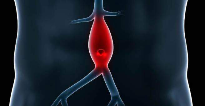
- Description: morbid outgrowth of a blood vessel, most commonly affected is the abdominal aorta, followed by thoracic and cerebral arteries, at rupture threaten life-threatening bleeding.
- symptoms: often symptom-free, depending on the situation but also pain, indigestion, cough, shortness of breath, headache, blurred vision or facial paralysis. When tearing extreme pain, circulatory collapse, coma
- Causes: Congenital malformations, familial predisposition, arteriosclerosis, hypertension, rarely bacterial infections
- Diagnosis: mostly incidental findings with abdominal ultrasound, brain scan or chest radiographs
- Treatment: Closure of the aneurysm, usually minimally invasive, by vascular prosthesis, stent, bypass, coiling, clipping, wrapping or trapping (see text). Smaller aneurysms often only need to be observed.
- Forecast: recognized in time, the forecast is good. If an aneurysm breaks, more than 50 percent of patients die.
Aneurysm: description
An aneurysm is a pathological weak spot of a blood vessel, at which the vessel wall widens usually bag-shaped, berry- or spindle-like. In most cases, aneurysms form in arteries because they have a higher pressure than in the veins.
Mostly the abdominal aorta is affected
In about 75 percent of the cases, the balloon-shaped Gefäßaussackungen occur in the course of the main artery (aorta) in the abdomen. But aortic aneurysms in the chest are common, as well as aneurysms in the brain and in the region of the popliteal fossa. Even an aneurysm in the heart is possible.
Often without complaints for a long time
Aneurysms often cause no discomfort. They are therefore often discovered only accidentally in an ultrasound scan or brain scan – or at worst only when they break. Then there is acute danger to life – because of the massive blood loss or in the head also by brain damage.
However, many people live with the vascular changes for decades without ever being told about it. Experts estimate that two to three percent of the adult population has an aneurysm.
What forms of aneurysms are there?
Depending on the type of vascular wall change, doctors distinguish the following types of aneurysms:
“True” aneurysm (aneurysm verum): In the so-called “true aneurysm”, the various layers in the blood vessel wall are all preserved throughout, but the vessel wall is bagged.
Split aneurysm (aneurysm dissecans): A layer in the wall of the blood vessel is torn and blood collects between the vessel wall layers.
“False” aneurysm (aneurysm spurium): This is not an aneurysm in the true sense. The aneurysm spurium has a leak in the vessel wall which is sealed from outside (e.g., by the surrounding tissue). Then, around the blood vessel, a hematoma forms, which transforms into connective tissue in the long term.
Aneurysm: symptoms
If an aneurysm is not too big, it usually does not make itself felt. Which symptoms cause larger specimens depends on their location.
Aortic aneurysm in the stomach: symptoms
If an aortic aneurysm of the abdominal aorta is so great that it presses on surrounding structures, the following symptoms may occur.
- Pain, especially in the lower abdomen, usually stinging and persistent, regardless of the body position
- Back pain that radiate into the legs
- rare indigestion
- palpable, pulsating structure under the abdominal wall
Burst aortic aneurysm in the abdomen
The larger the aneurysm, the greater the risk of a tear (rupture). This is especially true for aortic aneurysms with more than six centimeters in diameter.
If such an aortic aneurysm breaks, unbearable abdominal pains suddenly appear, radiating into the back. There are also nausea and nausea.
Due to the massive blood loss of the blood pressure drops quickly. The patient suffers a circulatory shock. Such bleeding is an absolute emergency! About half of those affected do not survive a burst aortic aneurysm.
Aortic aneurysm in the chest area: symptoms
If the aneurysm is located on the main artery at the level of the thorax (thoracic aortic aneurysm), the following symptoms may occur:
- chest pain
- to cough
- morbid breathing sound (stridor)
- hoarseness
- dysphagia
- Shortness of breath (dyspnoea)
If the airways are severely restricted in a thoracic aortic aneurysm, pneumonia can occur again and again.
Burst aortic aneurysm in the chest
Thoracic aneurysms over five and a half inches in diameter are particularly dangerous. If they break, they usually cause severe chest pain. The symptoms are similar to those of a heart attack. A rupture is fatal in three out of four cases.
Symptoms of an aneurysm in the brain
Some brain aneurysms (intracranial or cerebral aneurysm) press on individual neurons. Especially often the eyes are affected, also facial paralysis occur.
Bursted brain aneurysm
If the vascular wall ruptures in an aneurysm in the brain, massive symptoms appear. Most commonly, so-called subarachnoid hemorrhage, short SAB. It bleeds in the space between the brain and the meninges, more specifically the spider skin (arachnoid).
Due to the solid skullcap, the blood can not escape and quickly exerts increased pressure on the brain. The symptoms of an aneurysm in the brain occur due to increased intracranial pressure:
- Sudden onset, strongest headache
- nausea
- Vomit
- stiff neck
- dizziness
- drowsiness
- Unconsciousness or coma
If the patient survives, stroke-type consequential damage such as hemiplegia is possible.
Aneurysm symptoms in the popliteal fossa
An aneurysm of the popliteal artery (popliteal artery) usually goes unnoticed. However, if the popliteal aneurysm has a diameter of more than three centimeters, a blood clot (thrombosis) may form.
As a result, the lower leg is no longer sufficiently supplied with blood. Especially the calf hurts, there are emotional disorders such as tingling, numbness and cold feelings.
If the blood clot is carried along by the bloodstream, it can close a vessel at a narrower point, for example in the lung (pulmonary embolism).
Aneurysm: causes
Whether an aortic aneurysm or aneurysm in the brain – the vascular glands can be innate or develop only in the course of life. Unfavorable genetic predisposition play a role, but so does a lifestyle that is detrimental to the vessels.
Congenital malformation
Sometimes aneurysms occur frequently within a family, for example, due to a hereditary connective tissue weakness. For example, the risk of an aneurysm increases from one to two percent when an aneurysm is detected in a first-degree relative, and four percent when two close relatives are affected.
Also, genetic disorders such as Marfan syndrome or Ehlers-Danlos syndrome often cause aneurysms.
Many aneurysms also occur due to congenital malformations of the blood vessels, without there being a familial accumulation.
Higher age
The risk of an aortic aneurysm increases with age. The reason is that the structure of the vessel wall changes over the years. It is less elastic and can more reliably absorb the high pressure in the main artery. It develops weak points in the vessel wall, which eventually give way – an aneurysm develops.
high blood pressure
Another important risk factor is high blood pressure (hypertension). With every heartbeat, a blast of pressure rolls over the arteries of the body. If the blood exerts particularly high pressure from inside, it damages the vessel walls.
arteriosclerosis
In more than 50 percent of cases, vascular calcification (arteriosclerosis) is the cause of an aneurysm. Lime and fat deposits (plaques) on the vessel walls cause them to lose their elasticity. The vessels can absorb the pressure with increasing age getting worse.
Bacterial infections
Rarely, bacterial infections are involved in the development of an aneurysm – for example, in syphilis or tuberculosis. During the infection, the vessel wall becomes inflamed. Finally, a Gefäßaussackung arises. This is called mycotic aneurysm.
Mechanical injuries of the vessel walls
The most common cause of an aneurysm spurium – the “false aneurysm” – is injury to the vessel wall. For example, cardiac catheterization, which involves pushing a narrow, flexible tube across the leg artery to the heart, can damage the vessel wall.
Aneurysm: diagnosis
Doctors often discover an aneurysm as part of a routine examination, for example in the case of an abdominal ultrasound, X-ray of the lungs or brain scan. This can be an aneurysm recognize.
Even when listening with a stethoscope, the doctor sometimes detects suspicious flow noises over the vessel lining. In lean people, an abdominal aortic aneurysm with a diameter of more than five centimeters can usually be felt as a pulsating swelling through the abdominal wall.
Imaging procedures
Depending on the situation, details of the size and danger of an aneurysm can provide a cardiac ultrasound, a computed tomography (CT) or magnetic resonance imaging (MRI) and possibly an angiography (representation of the vessels). In the case of an aortic aneurysm in the breast section, a special ultrasound procedure is also used. The transducer is inserted over the esophagus (transesophageal ultrasonography).
Aneurysm: treatment
There are several procedures for treating an aneurysm. Whether one treats at all and if so how, depends on various factors:
- Size of the aneurysm
- location
- Probability of a rupture
- surgical risk
- Condition of the patient
- Desire of the patient
Aneurysm – operate or wait?
Smaller, asymptomatic aneurysms often do not need immediate treatment. She controls the doctor once a year, slightly larger twice a year by ultrasound. It is important that the blood pressure remains in the lower normal range (120/80 mmHg). For this purpose, a blood pressure lowering drug may be used.
If an aortic aneurysm reaches a diameter of six centimeters in the abdominal aorta or five and a half centimeters in the chest, the danger of the vessel wall tearing up increases. In this case, an aortic aneurysm must be treated. However, even during the procedure, there is a risk that the vessel will rupture.
With an aneurysm in the brain, the situation is often even more delicate. Depending on the location and condition of the vessel, there is a different degree of danger of causing a brain injury during surgery, which can have serious permanent damage. Surgery or not – this decision must be made individually by the doctor and the patient.
Surgical treatment of the aortic aneurysm
For larger aortic aneurysms in the chest or abdomen, two surgical methods are available:
Stent (endovascular procedure)
An aortic aneurysm can often be stabilized using a stent. Over a small incision in the inguinal artery, the doctor pushes a small tube to the wall of the wall. This bridges the vulnerability.
vascular prosthesis
In the operation of an aortic aneurysm, the surgeon cuts across the dilated portion of the arterial wall and replaces it with a tubular or Y-shaped vascular prosthesis.
If there is an enlargement close to the heart, the aortic valve often has to be replaced additionally (artificial flap).
Treatment brain aneurysm
For the treatment of an aneurysm in the brain, there are primarily two methods that complement one another: clipping or coiling. It depends in particular on the shape of the aneurysm, which method e is more promising individually.
coiling
When coiling, the doctor usually stabilizes the vessel with the help of a wire mesh (stent) and wears the aneurysm in the brain with special platinum spirals from the inside. To do this, he first pushes a microcatheter over the groin into the relevant cerebral artery. Although these microspirals fill the brain aneurysm only partially. However, there are platelets, which clump together and thus close the aneurysm.
Advantage of the method is the minimally invasive, gentler access to the aneurysm. However, it does not apply to broad-based aneurysms. Another disadvantage of the method is that it often does not completely occlude the aneurysms. They have to be checked regularly and if necessary treated again.
clipping
If coiling is not possible or if the aneurysm is already torn, it will usually be clipped. The surgeon closes the aneurysm in the brain with the help of a mini clip. He first opens the skull. It creates a gentle access to the vessel lining between the natural convolutions of the brain. The occlusion of the aneurysm is performed with the aid of a high-resolution surgical microscope.
With this method, the aneurysm can usually close reliably. Follow-up examinations are then no longer necessary. The procedure is less gentle than a coiling.
Wrapping
Another neurosurgical option is wrapping. It is used in complicated cases when clipping is not possible. The unstable vessel is stabilized from the outside by wrapping the vessel. This can be done with the help of the body’s own tissue or else with gauze or plastic. That’s why a connective tissue capsule forms.
trapping
Another method is the so-called trapping. In doing so, the aneurysm in the brain is relieved by placing clips or balloons in front of and behind it. However, the procedure is only possible if the affected cerebral artery is not the only supply path for certain areas of hearing.
Treatment of an aneurysm of knee artery
In an aneurysm of the popliteal artery, the treatment is usually performed by a bypass operation. The surgeon transplants a piece of a blood vessel that he has previously taken from another part of the body, bypassing the aneurysm.
Living with aneurysm
In addition to the stabilization of the vessel, it is crucial to consistently minimize the causes of the aneurysm. This is especially true for patients whose aneurysm is observed but not stabilized surgically.
- Main Action: Keep blood pressure low! This is usually done with the help of beta-blockers or ACE inhibitors
- balanced diet with low animal fats, high fiber and vegetables
- Sporting activity
- Avoidance or reduction of obesity
- Abstain from nicotine
- Little alcohol
Avoid blood pressure peaks!
Patients with untreated aneurysm should avoid sudden spikes in blood pressure that could provoke tearing.
- Avoid heavy lifting.
- Learn to breathe properly under exercise.
- Let asthma or chronic bronchitis be treated as best as possible. When coughing, the blood pressure increases!
- Avoid blockages and treat them if necessary. Even when pressing on the toilet, the blood pressure rises.
- Do not overdo it during exercise, especially during strength training.
- Strive for a serene attitude to life.
Aneurysm: disease course and prognosis
The prognosis of an aneurysm depends on several factors. The decisive factors are the diameter of the vessel lining and the speed with which it enlarges.
At rupture high risk of death
The most serious complication is the rupture of the aneurysm (rupture) – it can cause life-threatening bleeding. Mortality in such a case depends on where the aneurysm is located.
Thus, the mortality in a ruptured abdominal aortic aneurysm over 50 percent, ruptures the main artery in the chest, it is even up to 75 percent. If an aneurysm of a blood vessel tears in the head, about half of the patients die within the first 28 days. Others suffer lifelong damage typical of a stroke, including hemiplegia and reduced speech and vision.
If an aneurysm is discovered and treated in time, the chances of recovery are good. However, surgery, especially in the brain, involves its own risks.