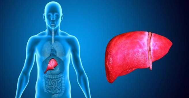
Liver metastases are lesions (daughter ulcers) in the liver. These usually emanate from other malignant tumors such as colon cancer. Liver metastases are usually painless, real symptoms appear very late. The settlements are best removed surgically. All important information about liver metastases can be found here.

Liver metastases: description
Liver metastases are – like all metastases – the dislocations of a malignant tumor that has its seat elsewhere (primary tumor) elsewhere in the body. The ability to spread to other parts of the body, that is to “metastasize”, is a hallmark of malignant tumors.
In liver metastases, a distinction is made between primary and secondary neoplasm of cancer cells in the liver. Under primary liver metastases one understands directly in the liver originated cancer cells for example from one hepatocellular carcinoma (Liver cell cancer) or one cholangiocellular carcinoma (Bile duct cancer). The secondary liver metastases are more common and arise from other malignant tumors that have spread to the liver. The liver, for example, is the organ that metastasizes in colon cancer patients in 75 percent of cases.
Liver metastases, depending on the nature of the original tumor, occur soon after the primary tumor develops, or only a few years later. If they are already present at the first diagnosis, doctors call that synchronous liver metastases, This is not uncommon, about 15 to 25 percent of colorectal cancer patients already have metastases at diagnosis. In contrast, there are still the metachronous liver metastases. They develop during the course of the cancer or after the original tumor has been removed. This is the case in about 20 to 50 percent of patients.
Liver metastases: symptoms
The symptoms of liver metastases are nonspecific and are therefore more noticeable in the later course of the disease for the person concerned. Weight loss, loss of appetite and general weakness may be the first signs of liver metastases. They are usually not painful. If the tumor is located centrally in the liver, this can manifest as a yellowing of the skin (jaundice, jaundice). It arises because the bile pigment is no longer sufficiently degraded by the liver and finally stored in the skin and in the whites of the eyes (sclera).
Liver metastases: causes and risk factors
A typical feature of a malignant tumor is that the tumor can grow into neighboring organs (infiltrative growth). Most importantly, the cancer cells spread to another location of the body, they metastasize. This can be done in two ways: thehaematogenous metastasis Degenerate cells detach from the tumor and circulate through the blood into other parts of the body. In colon cancer, for example, the tumor cells enter the bloodstream and flow downstream via the portal vein, first into the liver, where they attach themselves and continue to grow unchecked. Another way is thelymphoid metastasis: The tumor cells are transported via the lymphatic route to nearby lymph nodes. This distribution is rather rare in liver metastases.
In liver metastases, the original tumor is often located in the digestive tract. Therefore, they are especially common in gastric, oesophageal (esophageal) or colorectal (colorectal) carcinoma. But also a cancer of the pancreas (pancreatic carcinoma) or the bile ducts can lead to liver metastases. Liver metastases after breast cancer, lung cancer and thyroid cancer are rather rare.
Liver metastases: examinations and diagnosis
If you suspect that you have liver metastases, an internist or oncologist is the right person to contact. This will first ask you about your medical history (anamnese). He may ask you the following questions:
- Have you lost weight by accident lately?
- Do you have fever?
- Have you noticed any unusual yellowing of your skin lately?
- Are you suffering from a diagnosed cancer?
Afterwards, the physician also examines you physically using various methods. First, a ultrasound performed to look for possible liver metastases. In particular, the size and condition of the liver can be analyzed. In addition, enlarged lymph nodes are a possible sign of liver metastases.
For a more detailed picture, the Computed Tomography (CT) used. If the diagnosis is unclear, the cross-sectional images help to better assess the liver. Another diagnostic procedure is the magnetic resonance imaging For this purpose, a special contrast agent is administered before the examination, whereby certain changes become visible.
In some cases, liver metastases are one incidental findingwhich is detected during palpation or ultrasound examination of the abdomen. Then it is important to find and treat the original tumor as quickly as possible. If nobody is found, one speaks of a so-called CUP syndrome (cancer of unknown primary). In some cases, liver metastases can also be diagnosed with ultrasound-guided fine-needle biopsy or punch biopsy.
Liver metastases: treatment
Liver metastases have long been considered incurable. However, there is now enormous progress in the treatment of liver metastases. Patients with liver metastases should be well informed and referred to a general and visceral surgeon with good experience in liver metastasis surgery. Based on the previously performed examinations, the doctor will ultimately decide which therapy is eligible.
Surgical removal
Basically, the initial tumor from which the liver metastases originate is treated first. Single (solitary) liver metastases can be surgically removed without destroying much of the liver. In order to operate, it must be ensured that no other secondary tumors are present and that the liver remains functional after the operation. The goal is to get as much healthy liver tissue as possible. The liver also has the special property of regrowth. Up to four-fifths can be removed, provided the remainder of the liver is healthy.
Unfavorable for surgery is when one of the liver metastases is near a hepatic artery, or so many visible metastases have already formed, that there is a high probability that some of them are not yet visible.
The success of the treatment depends on the location of the liver metastases and the general condition of the patient. Only about 10 to 20 percent of sufferers with liver metastases are actually eligible for surgery. Therefore, different therapy methods have been developed for a wider range of treatments.
chemotherapy
Chemotherapy means to treat the tumor cytostatics be used. Cytostatic drugs are drugs that inhibit the growth of degenerate cells. In some cases, the liver is diffused with secondary tumors, making surgical removal of liver metastases extremely difficult. However, prior (neoadjuvant) chemotherapy may still allow surgery in some patients. First, the systemic chemotherapy used, this shows no effect, can alternatively regional chemotherapy be applied.
The aim of the treatment is to reduce the size of secondary tumors and to protect liver tissue. In about 12 to 38 percent of cases, surgery for initially inoperable liver metastases after chemotherapy is still possible. About 17 percent of the treated patients were even cured afterwards. Downstream chemotherapy (adjuvant chemotherapy) may reduce the likelihood of tumor recurrence after surgery.
The down side, however, is that upstream chemotherapy in some cases damages the still healthy liver tissue. It may, for example, a chemotherapy-associated steatosis hepatis, a fatty liver occur.
Systemic chemotherapy: If the liver metastases are treated with systemic chemotherapy, the patient receives the cytostatics via the arm vein. The injection can also be done through a port, which is a special applicator placed under the skin in the chest with access to a central vein. The chemotherapeutic agents (cytostatics) thus reach the cancerous cells via the bloodstream. The therapy is systemic, that is, it affects the entire body.
Regional chemotherapy: Regional chemotherapy for liver metastases, however, predominantly affects the affected area. For this purpose, a liver pump system under the skin, in the liver area, attached. The cytostatic drug can be injected via it and reaches directly into the hepatic artery and finally into the liver metastases. Regional chemotherapy therefore works exactly where the liver metastases are located. Disadvantage is the complex treatment and that undiscovered metastases, which are located outside the liver, can not be achieved.
Local therapy procedures
If surgery of the liver is associated with too high a risk, if chemotherapy is not effective, or if liver metastases recur after treatment, there are no other therapies available. A treatment with Laser, radioactivity and radio frequencies represent less invasive and uncomplicated therapy procedures for the patient. The tumor tissue is destroyed locally, while the surrounding healthy liver tissue is spared. In particular, laser-induced interstitial thermotherapy (LITT) and radiofrequency therapy (RFA) are widely used. Alternatively, the Cryotherapy or alcohol ablation therapy be used. In local therapy, it is possible to repeat the treatment if metastases recur.
Laser therapy (LITT): In laser-induced interstitial thermotherapy, liver metastases are laser-treated by a probe placed under the skin. The procedure is among the minimally invasive, that is, gentle treatments.
Under local anesthesia (local anesthesia) the liver is punctured using computed tomography or ultrasound. Subsequently, a laser probe is placed under the skin in the liver tissue. This laser light with a wavelength of 1064 nanometers is transmitted via a glass fiber to the tumor cells, which heats and destroys them. Magnetic resonance imaging (MRI) closely monitors and controls the treatment.
The laser therapy has the advantage that it can be repeated without problems if necessary and locally only really affects the tumor tissue. However, LITT only treats liver metastases with a diameter of up to five centimeters; in addition, the remaining overcooked mass would be too large for the body to break down.
Unfortunately, this treatment does not count as a statutory cash benefit and often has to be borne by yourself. The cost is between 3,500 to 7,000 euros.
Selective Internal Radiotherapy (SIRT): In Selective Internal Radiotherapy, liver metastases are treated by radioactivity. Yttrium-90, a radioactive material, is administered via a catheter that is advanced through the inguinal artery to the hepatic artery. The radioactive material closes the small vessels of the liver metastases, so that they can no longer be supplied and eventually perish.
SIRT can cause pain in the liver and upper abdomen. In addition, complaints may occur in other organs such as the lungs, bile or stomach, as the radioactive substance is transported on the bloodstream.
For treatment, patients must remain in hospital for about two days, as radioactive radiation is still delivered to the liver metastases during this time. The treatment is usually taken over by the health insurance company.
Radiofrequency Therapy (RFA): Radiofrequency therapy allows destruction of the tumor by means of radio frequencies. Using imaging techniques such as ultrasound, computed tomography or magnetic resonance imaging, a needle electrode is pushed into the center of the metastasis. An alternating current is given off via the electrode which generates frictional heat through a high voltage and current density which overcooks the liver metastases. Up to a diameter of five centimeters, the liver metastases can be removed. The treatment takes about an hour.
cryotherapy: Cryotherapy is not treated with heat, but with cold. Using a probe, the liquid nitrogen is applied, temperatures up to minus 180 ° C can be achieved. The cells of the liver metastases are thus destroyed. This treatment is usually used for liver metastases, which are very large and therefore can not be operated on.
Alkoholablationstherapie: Alcohol ablation therapy injects pure alcohol into liver metastasis. The alcohol kills the cancer cells. However, this procedure is only used in primary liver cancer. In secondary liver metastases, only a portion of the cancer cells can usually be destroyed – the likelihood of recurrent liver metastases is therefore very high.
Liver metastases: disease course and prognosis
With complete removal of liver metastasis, the prognosis is very good for the patient. Of course, the prognosis depends essentially on the size and spread of the liver metastases and on the extent to which the original intestinal tumor has progressed. Median survival in patients with untreated liver metastases is 31 percent at one year, eight percent at two years, and three percent at three years. If colon cancer is the cause of liver metastases, the mean survival time is about four to 21 months. Will it succeed? liver metastases remove completely and at a safe distance, so that there is still enough healthy liver tissue, the chances of survival are up to 50 percent.